


P. austrocedrae - necrotic lesion in phloem
Photo Gallery
Site will be retired 9/1/2026
This site is no longer being developed and will be retired on September 1, 2026. Please contact us if you have any questions or would like to provide support to continue the project.
|
P. austrocedrae necrotic lesion in phloem 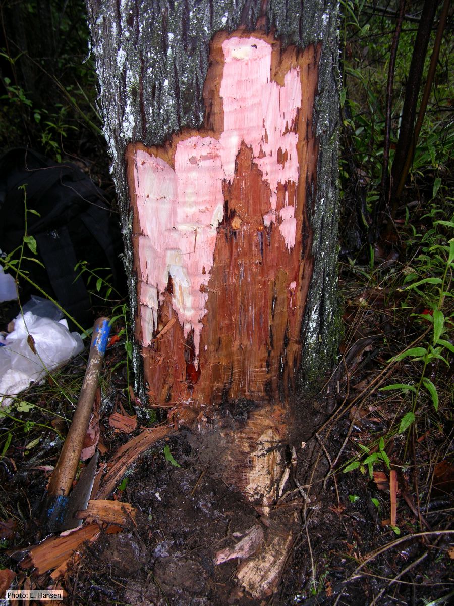 |
P. austrocedrae irregular sporangium 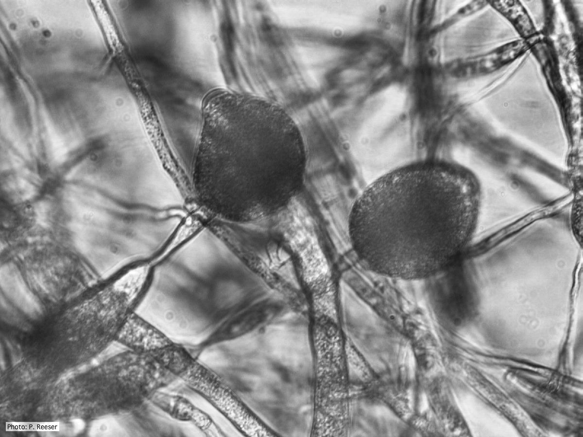 P. austrocedrae - irregular sporangium with lateral attachment and swelling in sporangiophore |
Mal del ciprés, dead and dying trees along river 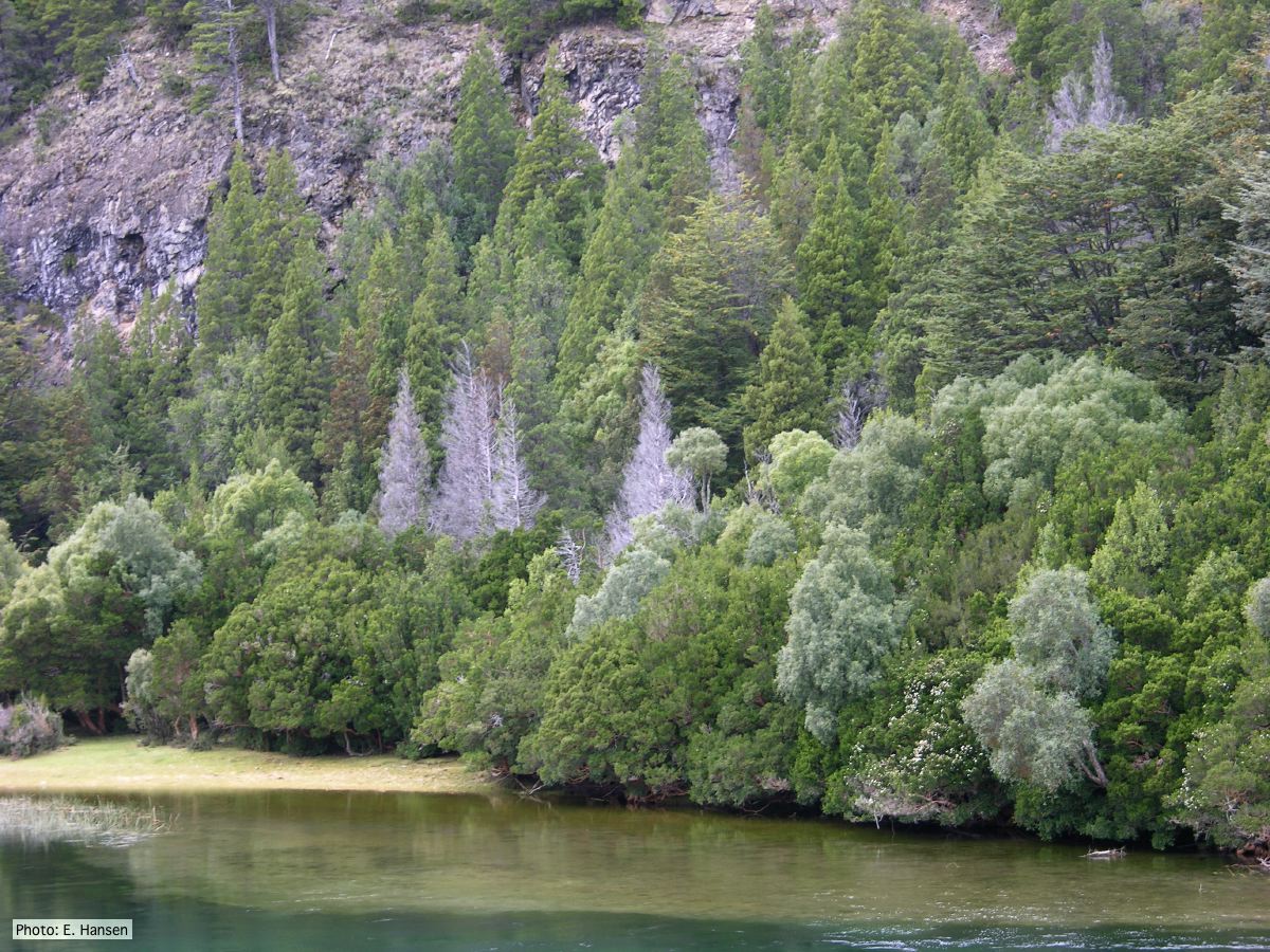 Mal del ciprés, dead and dying trees along river |
|
P. austrocedrae colony morphology on PDA  Colony morphology of P. austrocedrae at 16 C after four weeks on PDA |
P. austrocedrae oogonium 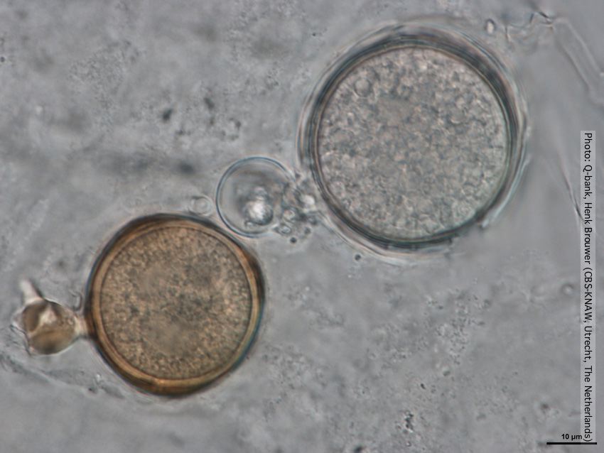 Oogonia with and without brown pigment, photo from Q-bank, used with permission |
P. austrocedrae colony morphology on Tomato juice agar with B sitosterol 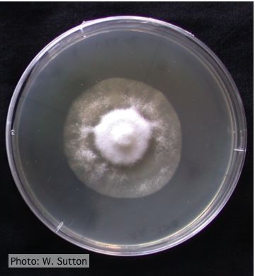 Colony morphology of P. austrocedrae at 16 ºC after 4 weeks on Tomato juice agar with B sitosterol |
|
P. austrocedrae - necrotic lesion in phloem 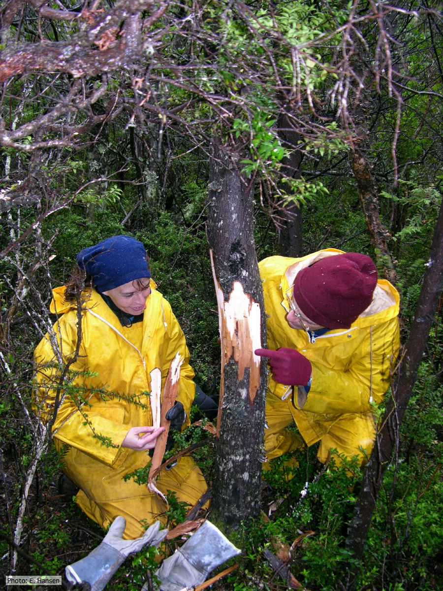 |
Austrocedrus associated with Mal del ciprés.  Austrocedrus with basal resin flow associated with Mal del ciprés. |
P. austrocedrae - hyphal swellings  Morphology of hyphae of Phytophthora austrocedrae, from Greslebin et al. 2007 |
|
P. austrocedrae - Mal del ciprés, stages of decline 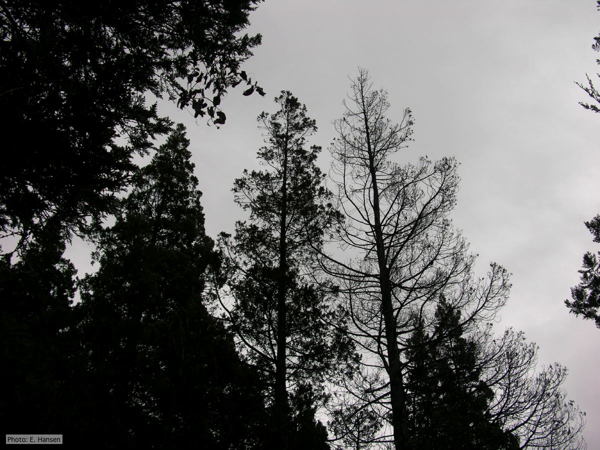 Mal del ciprés, stages of decline |
Necrotic lesion in phloem caused by P. austrocedrae 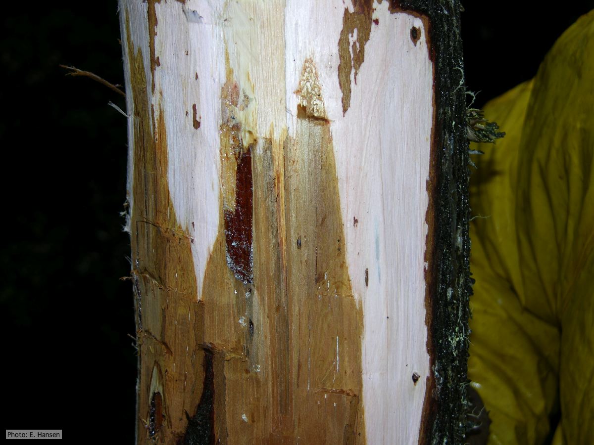 Necrotic lesion in phloem with resin pocket caused by P. austrocedrae |
P. austrocedrae oogonia drawing  P. austrocedrae. Morphology of oogonia, oospores and antheridia. Bar: 10 mm. Greslebin et al. 2007 |