

P. palmivora colony morphology on PDA
Photo Gallery
|
P. palmivora colony morphology on PDA 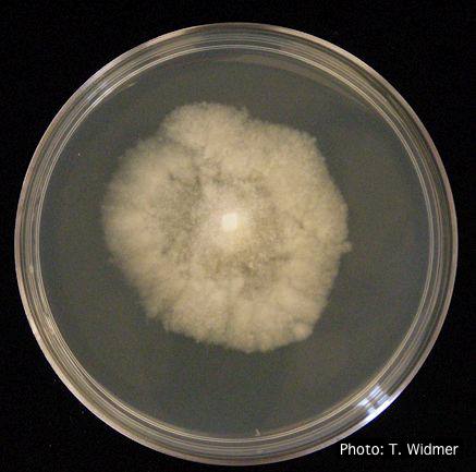 |
P. cinnamomi hyphal swelling 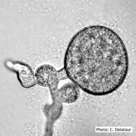 P. cinnamomi hyphal swelling (or thin walled chlamydospores) |
P. lateralis on Port Orford cedar 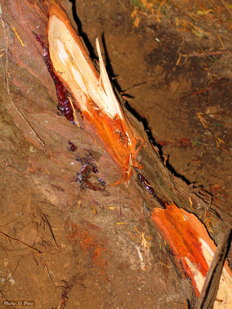 Small root lesions on Chaemacyparis lawsoniana |
|
Necrotic lesion in phloem caused by P. austrocedrae  Necrotic lesion in phloem with resin pocket caused by P. austrocedrae |
P. cambivora symptoms  Bleeding canker, European beech, OSU campus |
P. pluvialis on Pinus radiata needle  Clusters of sporangia emerge from stomata of an infected radiata pine needle. |
|
P. tentaculata chlamydospore  P. tentaculata chlamydospore with short hyphal projection |
P. cambivora symptoms 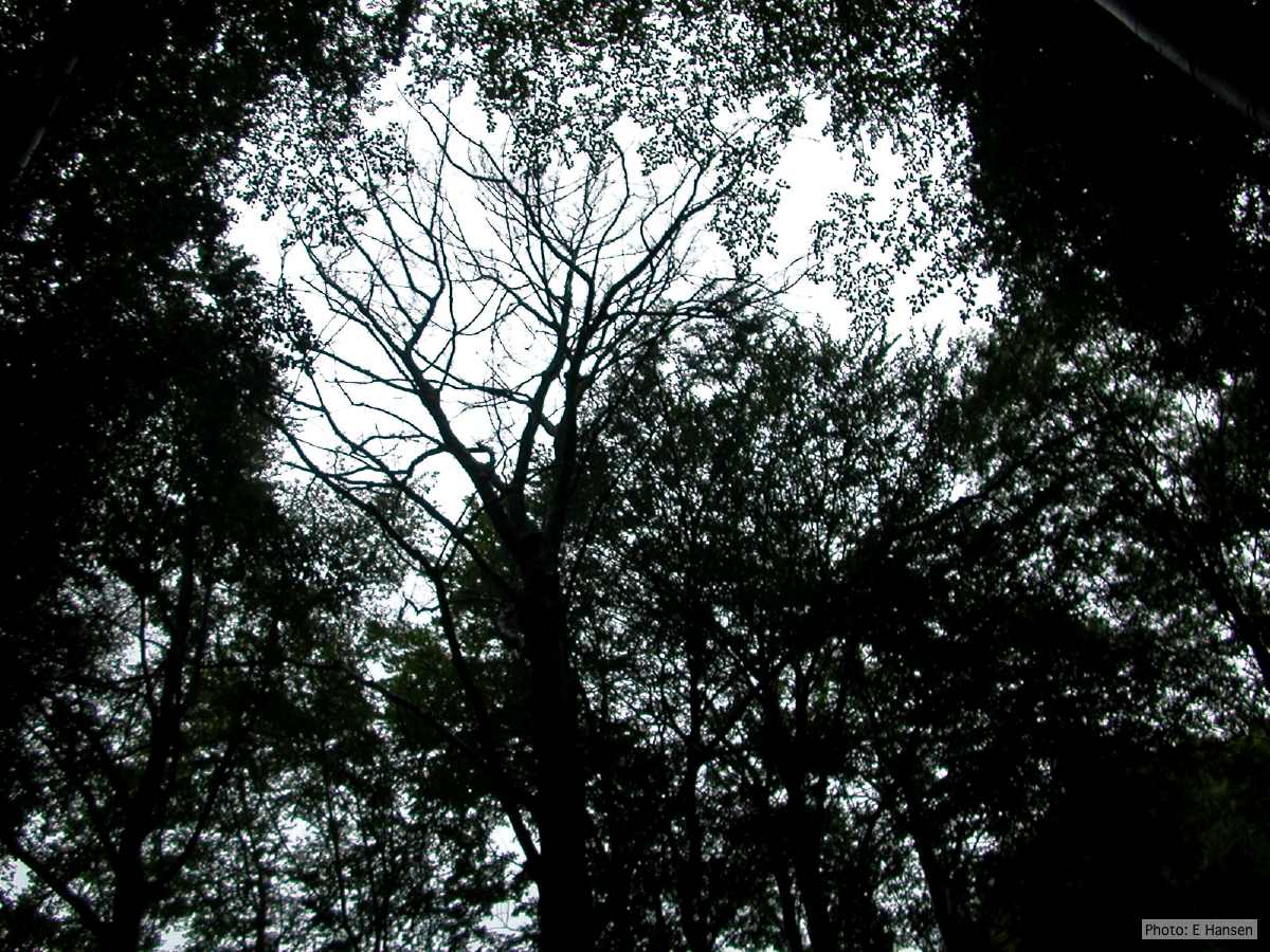 Dead beech in Germany |
P. frigida sporangia  Noncaducous sporangia showing ovoid shape and papillate condition |
|
P. cinnamomi in Australia  Death of Xanthoria and other native vegetation, Victoria, Australia |
P. cambivora inactive lesion on chinquapin  Inactive lesion of P. cambivora on chinquapin |
P. kernoviae leaf wilt 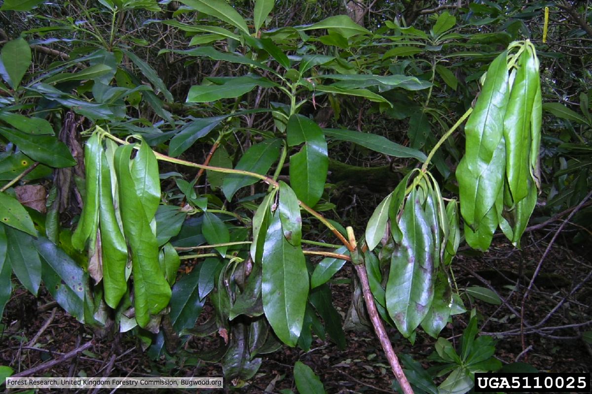 Wilted leaf of infected rhododendron |