

Oospores and oogonia with mostly paragynous but some amphigynous antheridia of P. tentaculata
Photo Gallery
|
P. tentaculata oogonia and antheridia 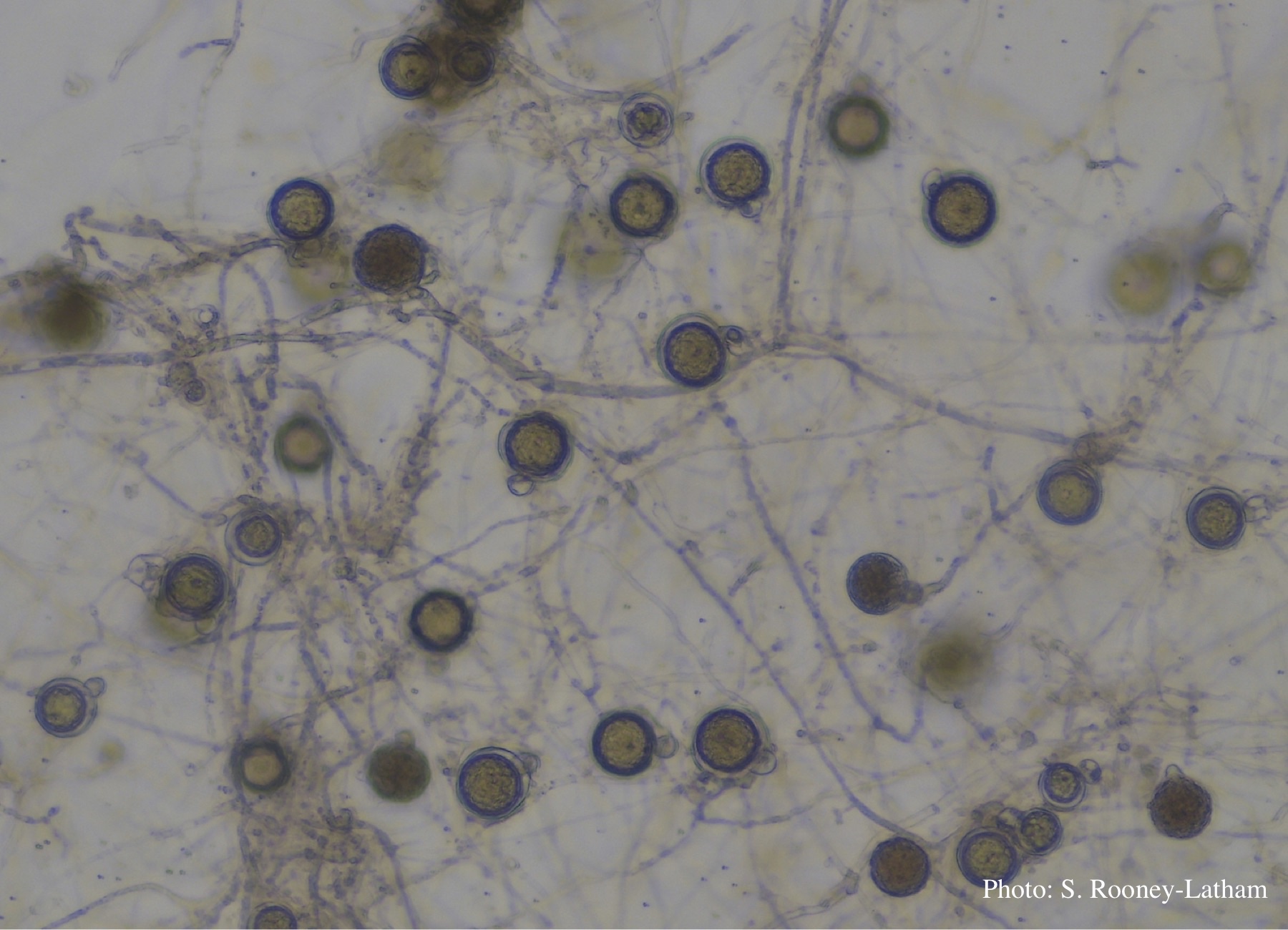 |
P. pluvialis oogonium and antheridium 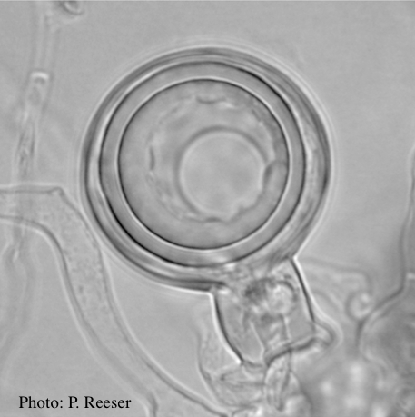 Oogonium and oospore with amphigynous antheridium |
P. pluvialis on Pinus radiata in New Zealand 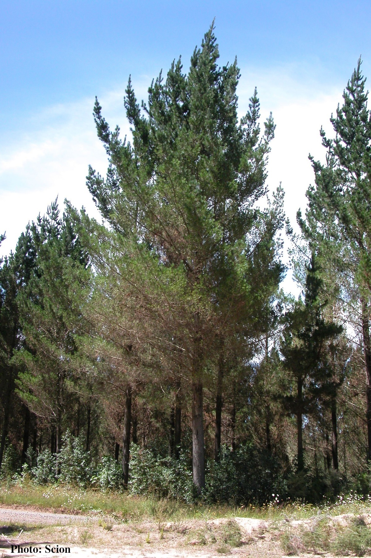 A stand of Pinus radiata trees affected by red needle cast disease. Note that frequently only the lower part of the crown is affected. |
|
P. ramorum chlamydospores 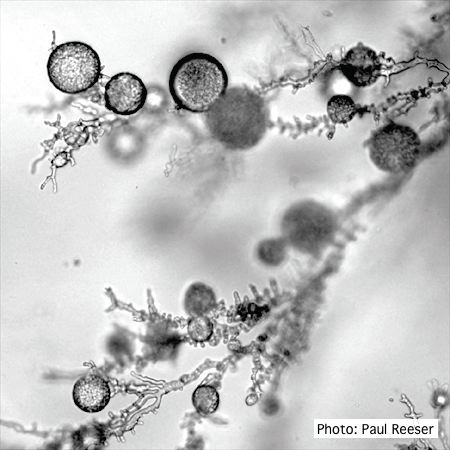 P. ramorum chlamydospores |
Growth of P. arenaria on half-strength PDA 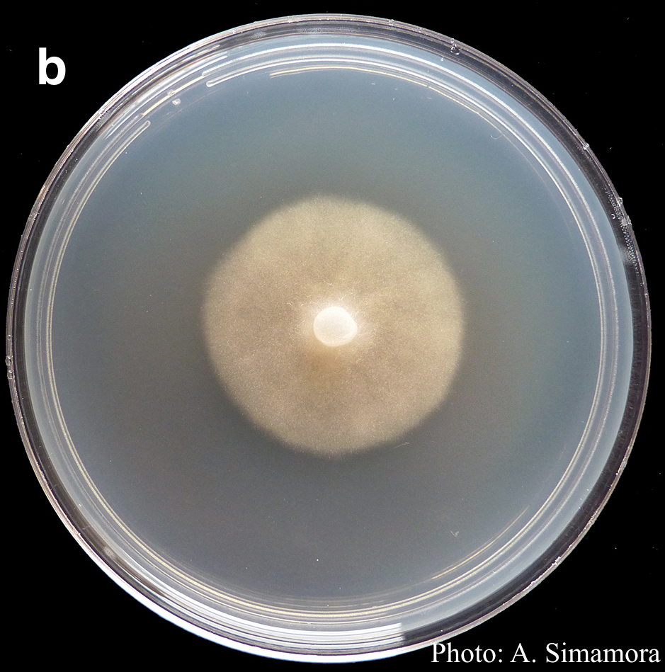 Colony morphology of Phytophthora arenaria after 7 days at 20°C on half-strength PDA |
P. cambivora coralloid hyphae 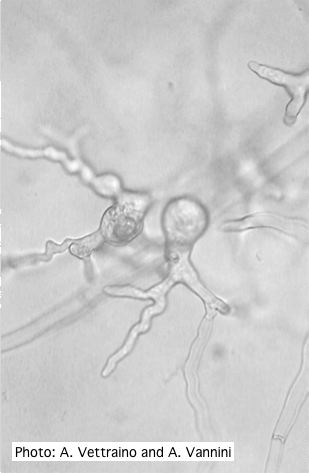 Coralloid hyphae with hyphal swelling-like structures |
|
P. palmivora sporangia
 P. palmivora caducous papillate sporangia
|
P. cinnamomi hyphal swellings 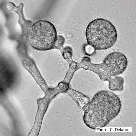 P. cinnamomi hyphal swellings (or thin walled chlamydospores) |
P. pinifolia sporangium 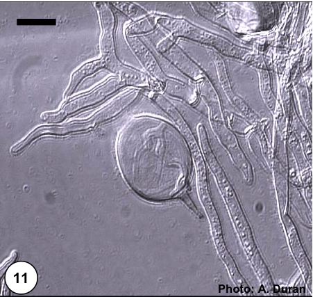 Non- papillate and caducous sporangia of Phytophthora pinifolia isolated from the infected P. radiata needles. |
|
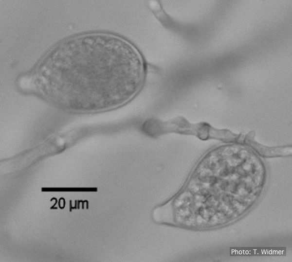
P. arenaria oogonium  Aplerotic oogonia of P. arenaria with paragynous antheridia. Scale bar = 20 μm |
P. ramorum sporangia 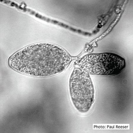 P. ramorum sporangia |
Chlamydospore of P. lateralis 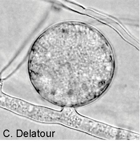 Terminal chlamydospore on a short side stalk |