


Sporangia of P. lateralis showing internal and external proliferation
Photo Gallery
Site will be retired 9/1/2026
This site is no longer being developed and will be retired on September 1, 2026. Please contact us if you have any questions or would like to provide support to continue the project.
|
P. lateralis sporangia 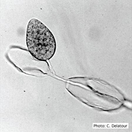 |
P. cambivora colony morphology on V8 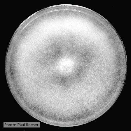 Colony morphology on V8 at 14 days |
Dead Port Orford Cedar trees 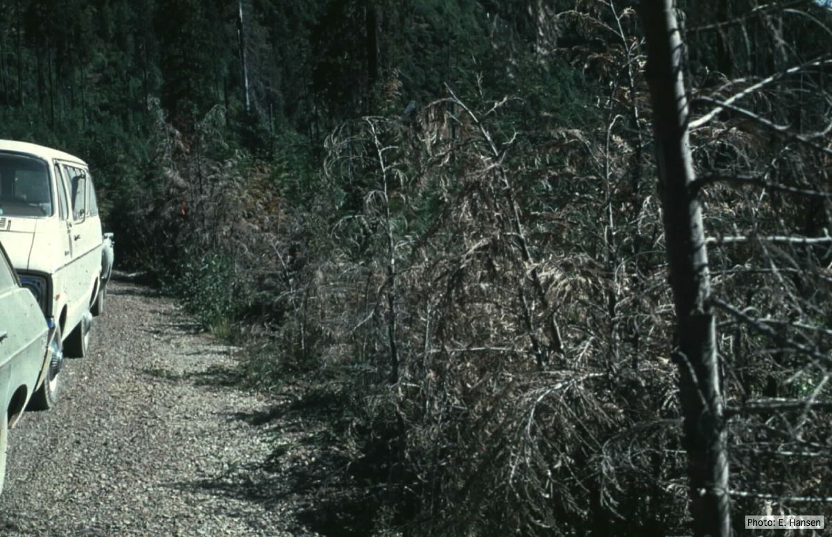 Dead Chamaecyparis lawsoniana trees along road |
|
P. cambivora sporangium with internal extended proliferation 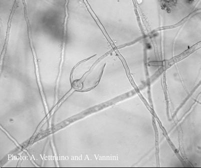 Empty sporangia showing internal extended proliferation |
P. agathadicida disease symptom 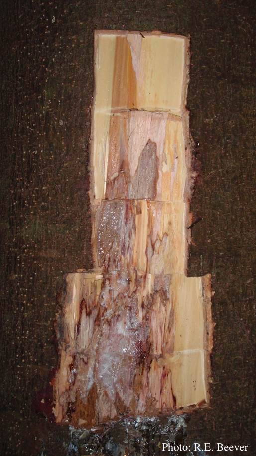 Excavated lesion, with outer bark removed showing extent of disease-front |
P. tentaculata sporangium 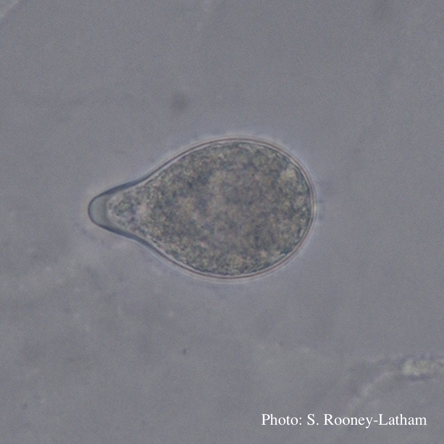 Papillate sporangium of P. tentaculata with an elongated neck or beak. |
|
P. katsurae sporangia 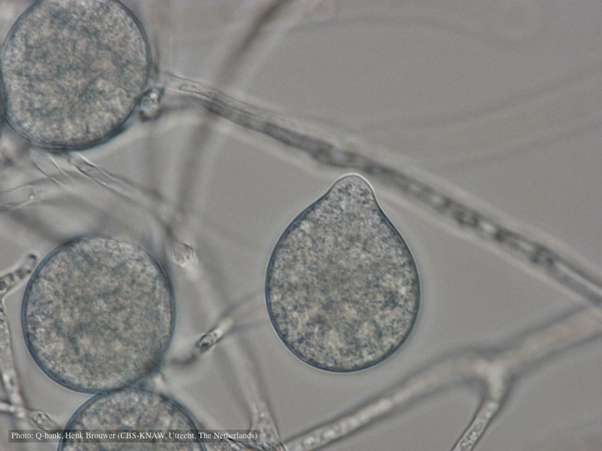 Papillate, non-caducous sporangia; photo used with permission from Q-bank |
P. cactorum bleeding canker  Bleeding canker on European beech (Fagus sylvatica) |
P. nemorosa colony morphology on V8 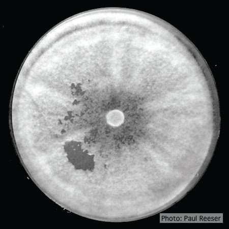 Colony morphology on V8 at 14 days |
|
P. pinifolia on Pinus radiata  Pinus radiata dead needles caused by DFP with healthy new growth from DFP |
P. cambivora colony morphology on MA 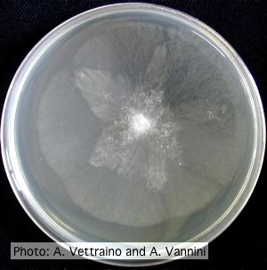 Appressed stellate colony morphology at 14 days at 20°C on MA |
P. katsurae disease symptoms 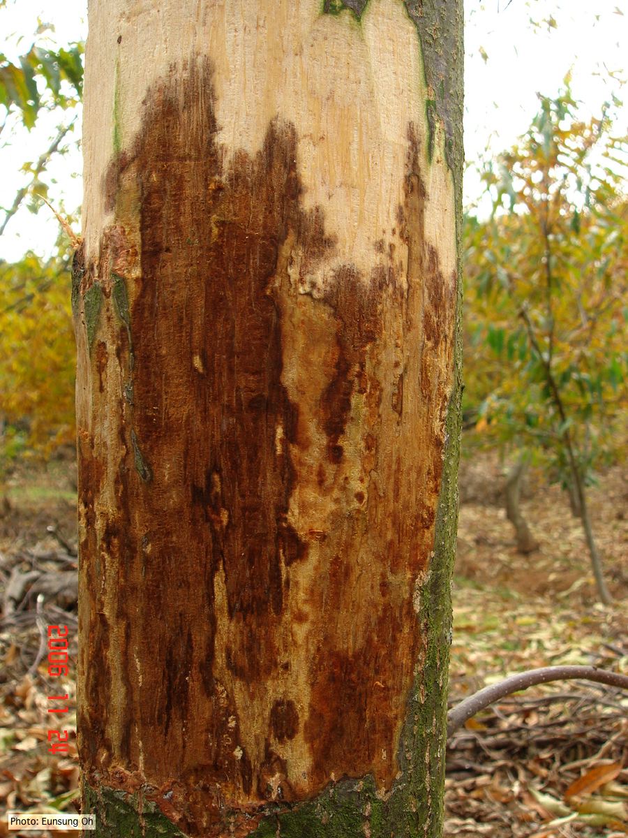 Infected chestnut (Castanea) with girdling canker |