

Sporangium showing ovoid and ovoid to spherical shape and papillate condition
Photo Gallery
|
P. boehmeriae sporangium 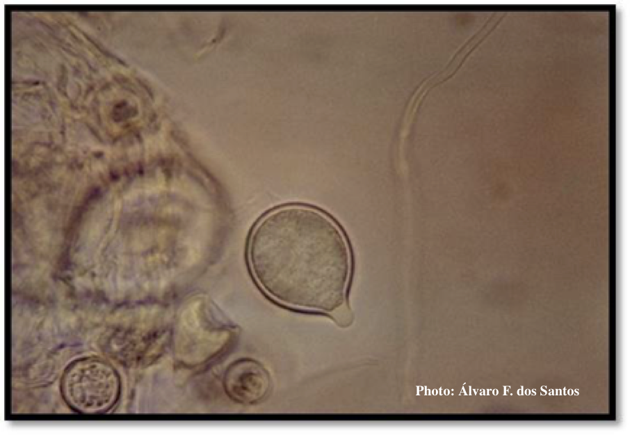 |
Growth of P. megakarya on V8 agar 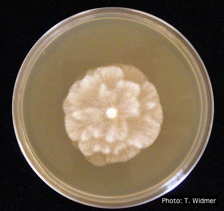 Growth of P. megakarya on V8 agar |
P. pluvialis colony morphology on carrot agar  Colony morphology on carrot agar at 20 days |
|
P. nemorosa colony morphology on PDA  Colony morphology on PDA at 14 days |
P. pluvialis symptoms on Douglas-fir 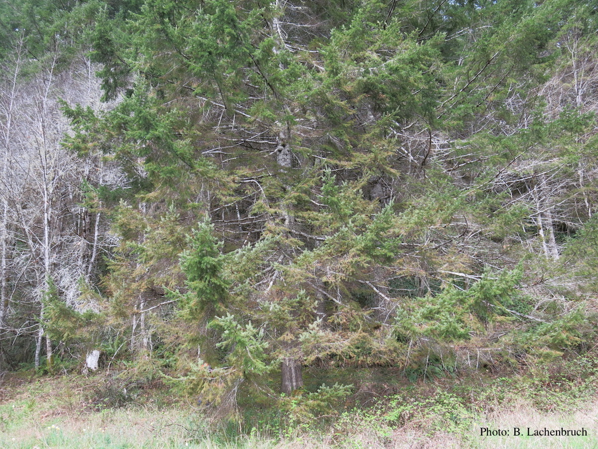 Red needle cast symptoms on Douglas-fir in western Oregon, 2015 |
P. cambivora colony morphology on PDA 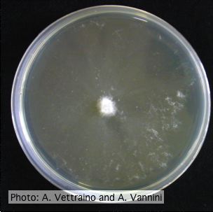 Appressed colony morphology at 14 days at 20°C on PDA |
|
P. pinifolia on Pinus radiata 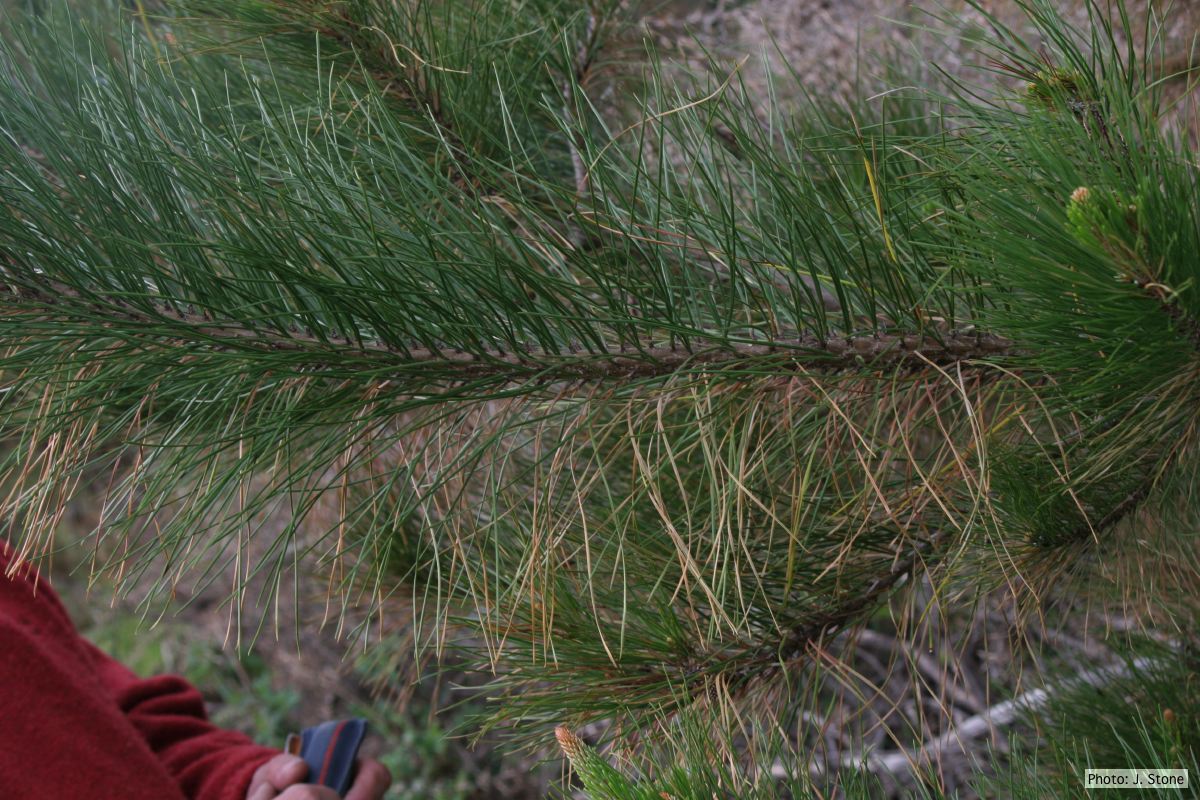 Dead needles on lower side of P. radiata branch. |
P. cambivora sporangium with internal extended proliferation  Empty sporagia showing internal nested and extended proliferation |
P. siskiyouensis colony morphology on V8 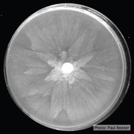 Colony morphology on V8 at 14 days |
|
P. cactorum bleeding canker 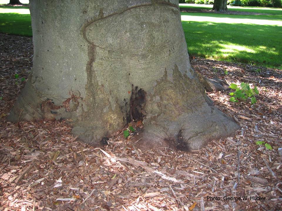 Bleeding canker on European beech (Fagus sylvatica) |
P. pinifolia on Pinus radiata 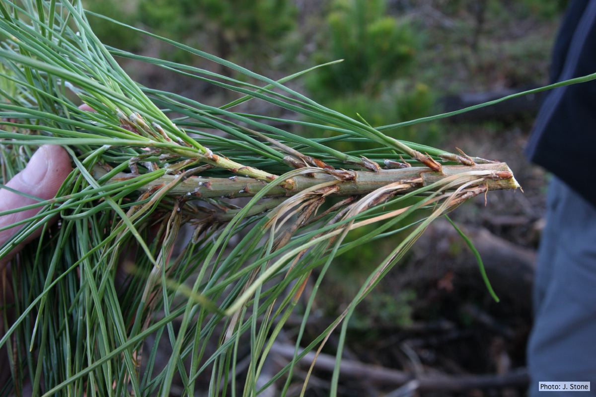 Pinus radiata, note grey and collapsed needle bases |
Comparative gametangial morphology of Phytophthora Clade 5 species  Comparative gametangial morphology of Phytophthora Clade 5 species, with SEM (top) and light microscopy (bottom). P. heveae has smooth walled oogonia with funnel-shaped, amphigynous antheridia. P. agathidicida has mildly stipulate oogonia with globose amphigynous antheridia. P.cocois has mildly bullate oogonia with reflexed amphigynous antheridia. P. castaneae has coarsely bullate oogonium with rugose protuberances and narrow amphigynous antheridia (Weir et al. 2015). |