

P. cinnamomi hyphal swelling (or thin walled chlamydospores)
Photo Gallery
Site will be retired 9/1/2026
This site is no longer being developed and will be retired on September 1, 2026. Please contact us if you have any questions or would like to provide support to continue the project.
|
P. cinnamomi hyphal swelling 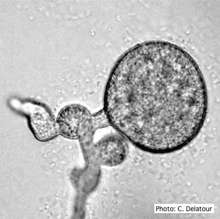 |
P. lateralis on Port Orford cedar 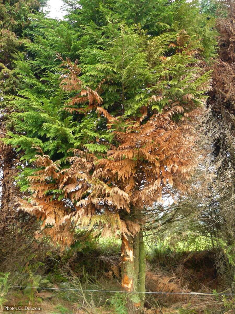 Localized branch infection of Chaemacyparis lawsoniana in Lopérec, France |
P. agathidicida oospores in planta  Oospores in the roots of kauri seedlings inoculated with P. agathidicida. The root has been cleared with potassium hydroxide and bleached with peroxide before being stained with trypan blue (scale bar =100 µm). |
|
Phytophthora chlamydospora sporangium 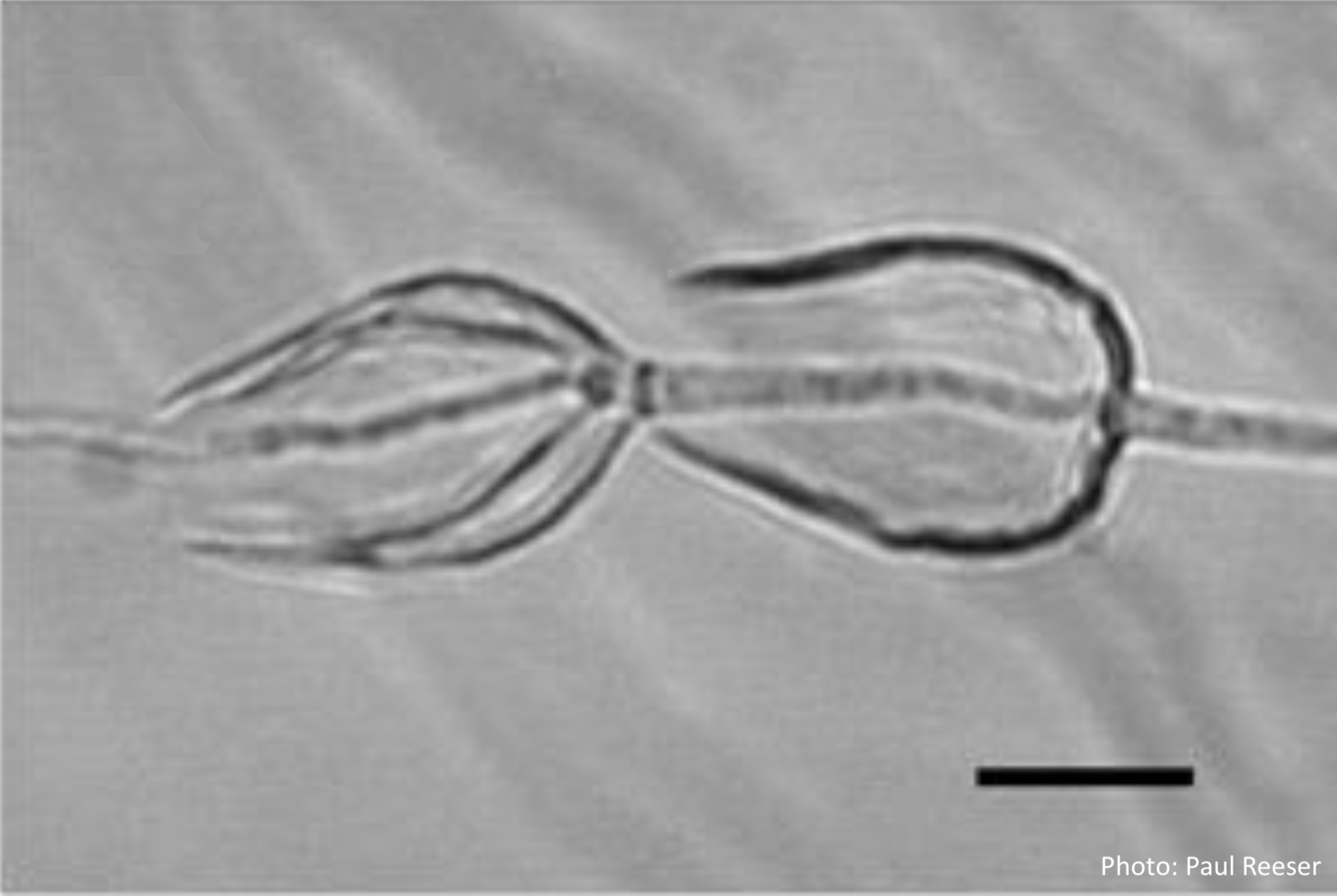 Phytophthora chlamydospora sporangia in water, showing internal proliferation. Bar is 20 µm. |
P. siskiyouensis oogonium with paragynous antheridium  P. siskiyouensis oogonium with paragynous antheridium |
P. pinifolia sporangia 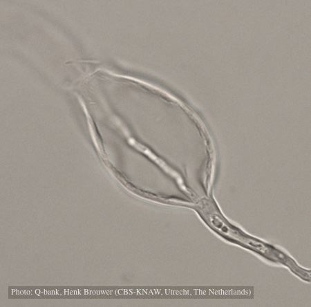 Sporangium with internal proliferation, photo from Q-bank, used with permission. |
|
P. pluvialis on Pinus radiata 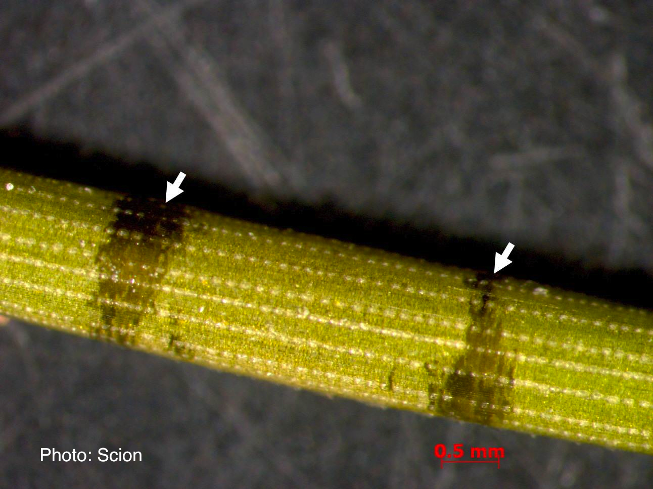 A Pinus radiata needle showing black resinous bands or marks consistent with the presence of red needle cast disease. |
P. cactorum bleeding canker 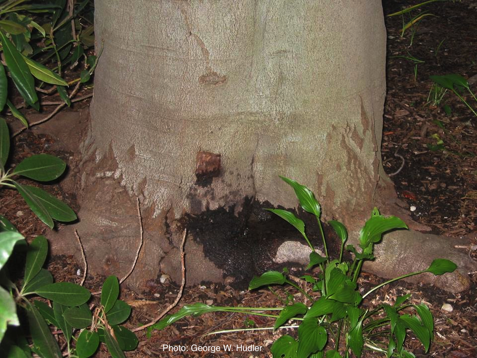 Bleeding canker on European beech (Fagus sylvatica) |
P. palmivora colony morphology on PDA 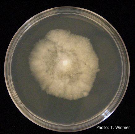 P. palmivora colony morphology on PDA |
|
P. cinnamomi sporangium 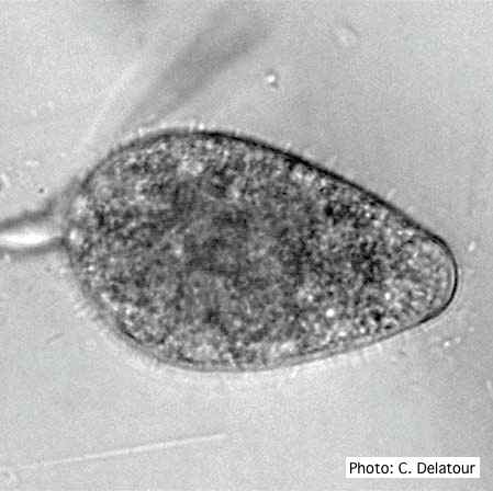 P. cinnamomi sporangium |
P. cambivora colony morphology on MA 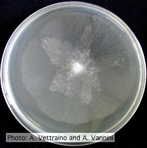 Appressed stellate colony morphology at 14 days at 20°C on MA |
P. nicotianae chlamydospore 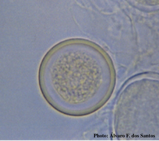 Globose chlamydospore (Fitopatol. bras. 2005) |