
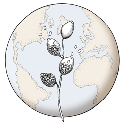

‘Blistered’ hyphal swellings in agar
Photo Gallery
Site will be retired 9/1/2026
This site is no longer being developed and will be retired on September 1, 2026. Please contact us if you have any questions or would like to provide support to continue the project.
|
P. nemorosa hyphal swellings 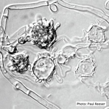 |
P. pinifolia hyphal growth 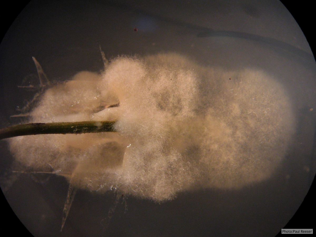 P. pinifolia pathogen growing from infected needle on selective agar |
P. siskiyouensis bleeding canker  Bole lesions in the tissues under the bark of a bleeding canker: discoloration in the secondary phloem tissue |
|
P. austrocedrae - Mal del ciprés, stages of decline 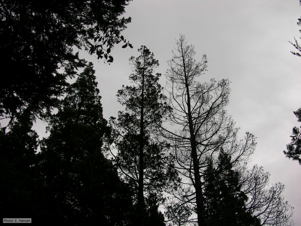 Mal del ciprés, stages of decline |
P. chlamydospora colony morphology on carrot agar 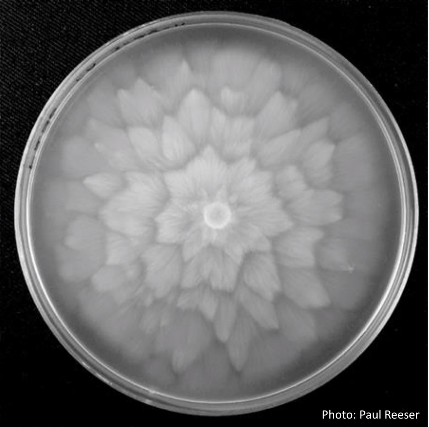 P. chlamydospora colony morphology on carrot agar |
P. pinifolia on Pinus radiata 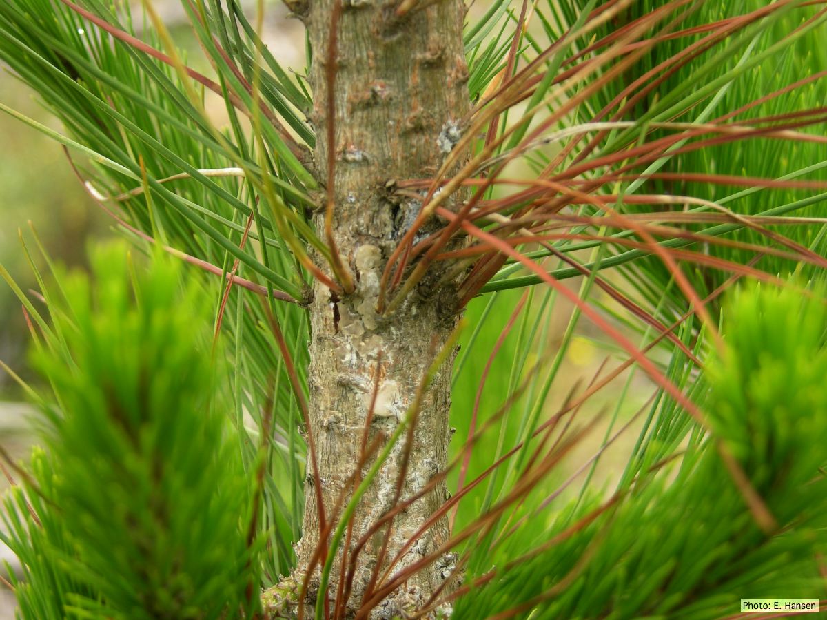 Pinus radiata, note Stem canker associated with necrotic needles. |
|
P. megasperma sporangia 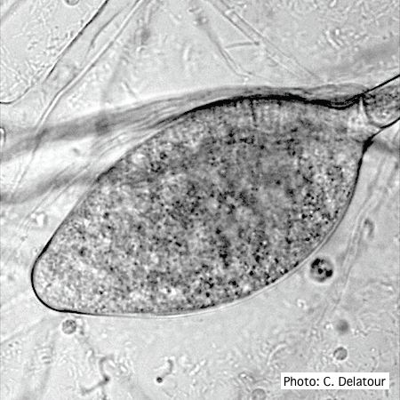 Ovoid, non-papillate sporangia showing internal proliferation of sporangiophore |
P. pluvialis sporangium 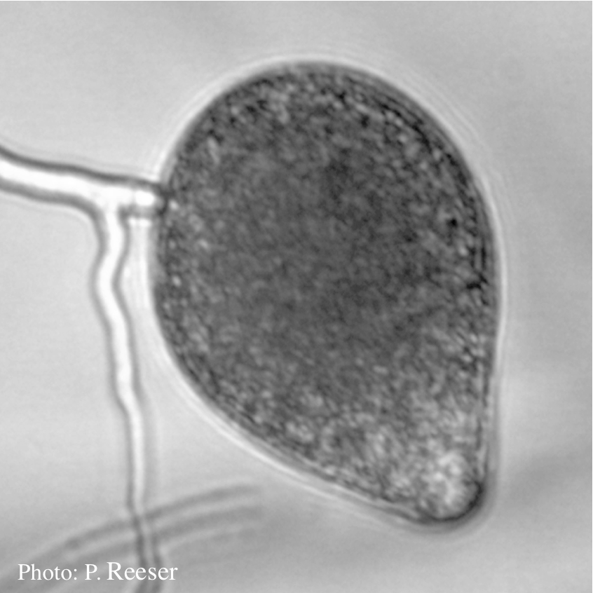 Sporangia showing typical ovoid shape and semi-papillate condition |
P. boehmeriae oogonia  Oogonia and oospores with amphigynous antheridia |
|
P. lateralis on Port Orford cedar 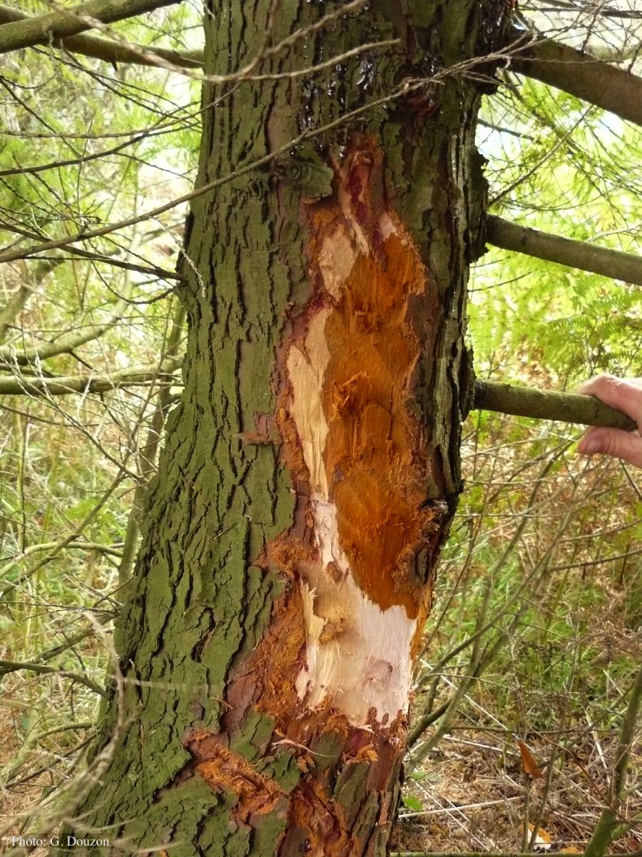 Bole lesion on Chaemacyparis lawsoniana in Lopérec, France |
Basal canker on Port Orford Cedar stump  |
P. tentaculata chlamydospore  P. tentaculata chlamydospore with short hyphal projection |