

Mortality of Juglans regia caused by Phytophthora cactorum.
Photo Gallery
Site will be retired 9/1/2026
This site is no longer being developed and will be retired on September 1, 2026. Please contact us if you have any questions or would like to provide support to continue the project.
|
Phytophthora cactorum disease symptoms on English walnut 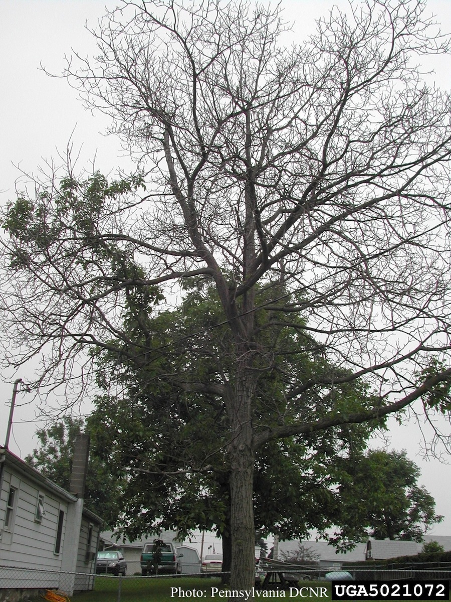 |
P. pinifolia sporulation 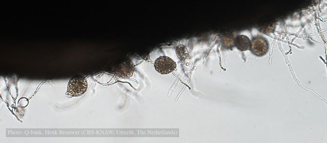 Sporulation on edge of hemp seed, photo from Q-bank, used with permission. |
P. palmivora colony morphology on V8 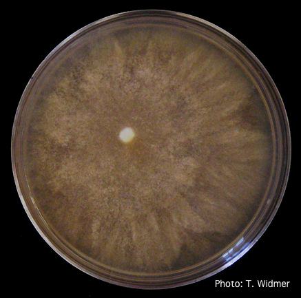 Growth of P. palmivora on V8 agar |
|
P. tentaculata disease symptoms on California mugwort 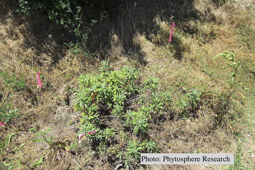 Outplanted California mugwort (Artemisia douglasiana) infected with P. tentaculata, 4.5 years after planting. Plant shows stunting and chlorosis. (P. cryptogea and P. lacustris were also baited from roots/soil of this plant). |
P. agathidicia sporangia 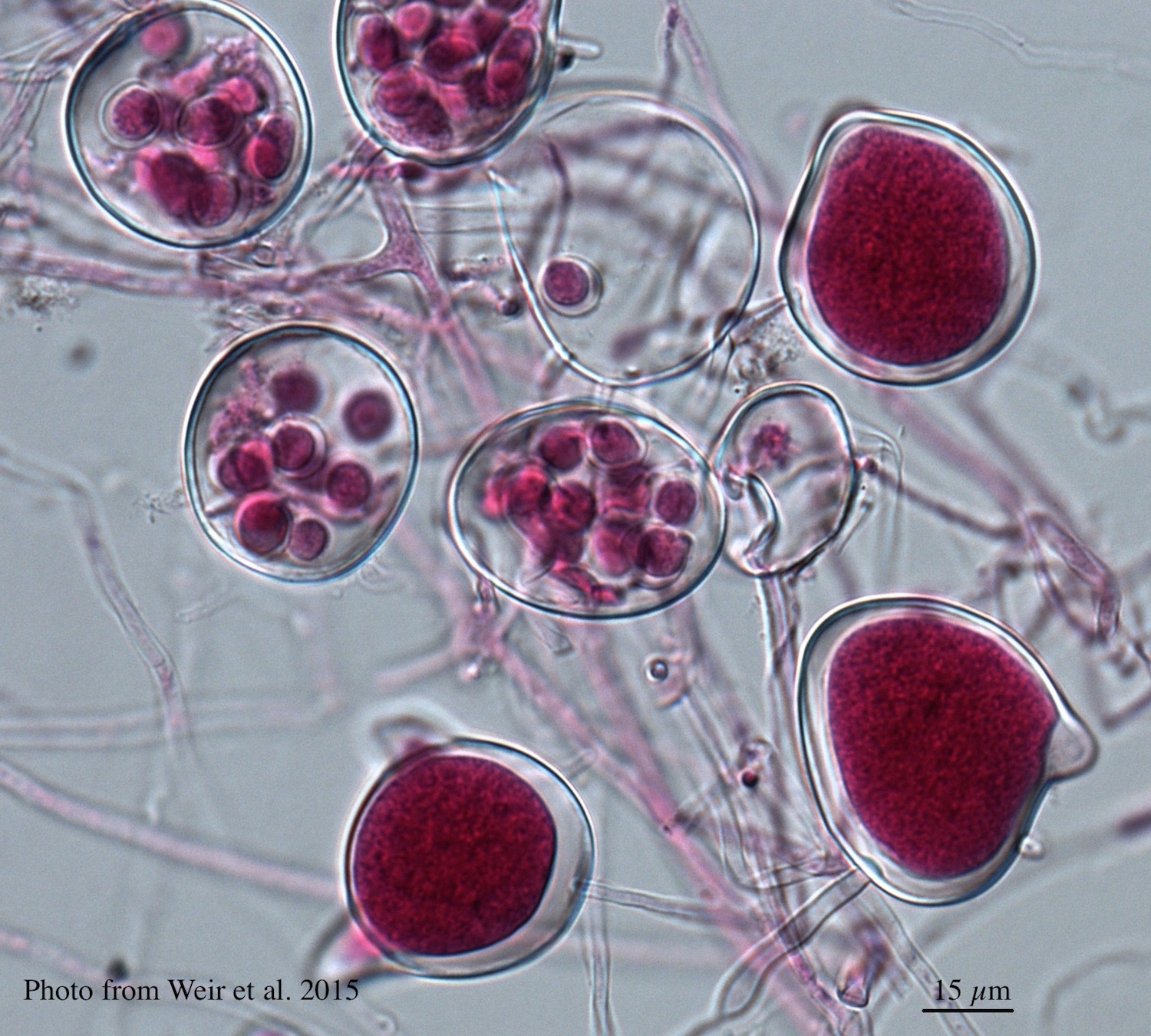 Differentiation of the cytoplasm within papillate sporangia into acid fuchsin stained zoospores |
P. kernoviae oogonia 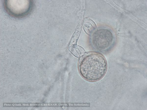 Oogonium with amphigynous antheridia, photo from Q-bank, used with permission |
|
P. kernoviae oogonia 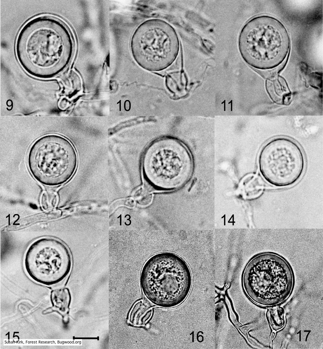 Mycol.Res 109, 853-859; Representative oogonia, antheridia and thick walled plerotic oospores of Phytophthora kernoviae. |
P. agathidicida lesion on kauri tree 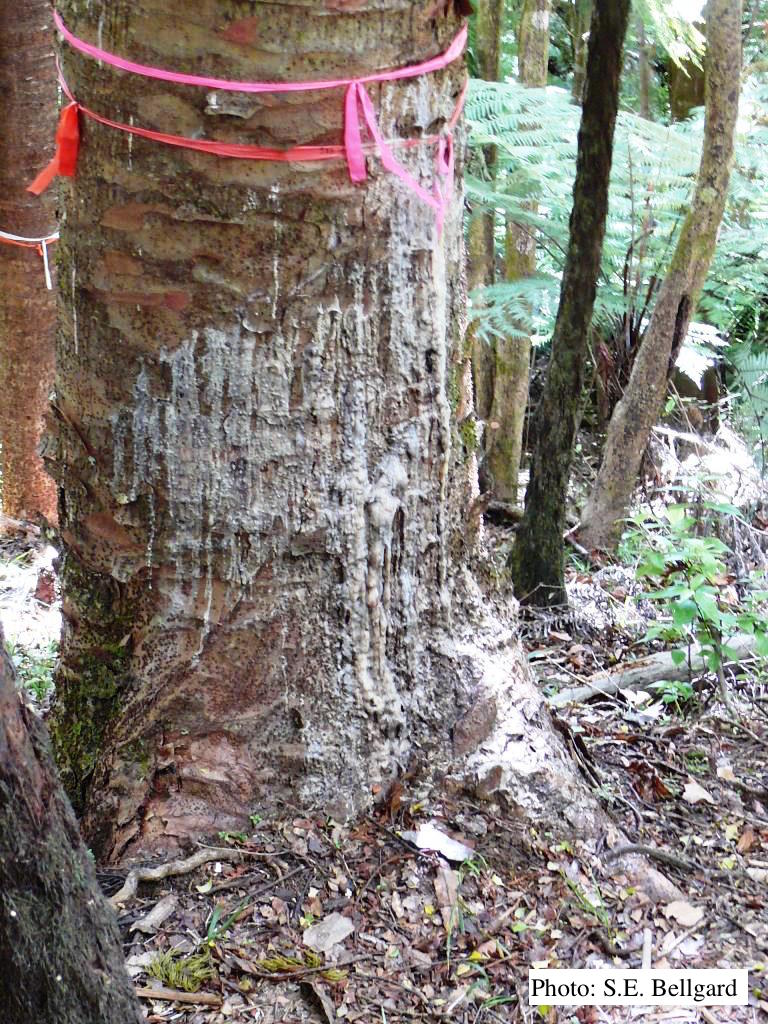 Advancing triangular lesion extending up the trunk of an 80 cm DBH kauri tree in the Huia Dam Site along Twin Peaks Track, Waitakere Regional Park |
P. nemorosa colony morphology on V8 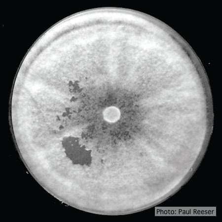 Colony morphology on V8 at 14 days |
|
P. alni oogonia 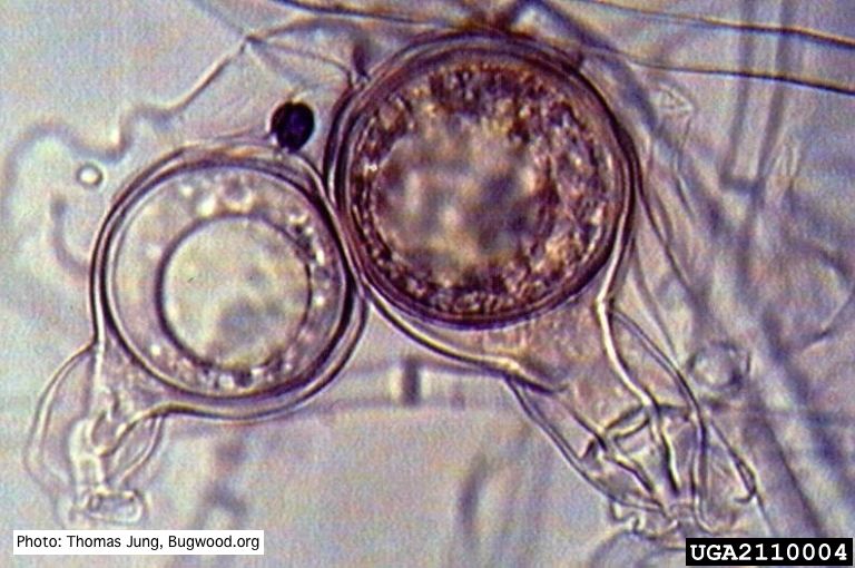 Smooth-walled oogonium of P. alni (Swedish variant) with oospore and amphigynous antheridium. |
P. nicotianae oogonia 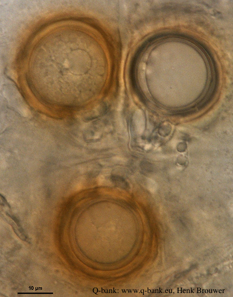 P. nicotianae oogonia 100x. Photo from Q-bank: www.q-bank.eu, Henk Brouwer (CBS-KNAW, Utrecht, The Netherlands) |
P. siskiyouensis bleeding canker 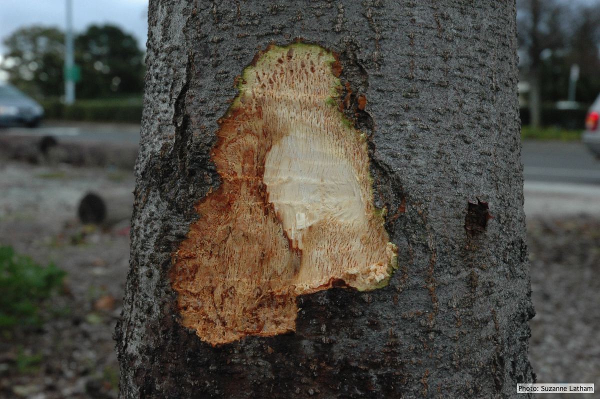 Close-up of margin area of bole lesions under the bark of a bleeding canker |