
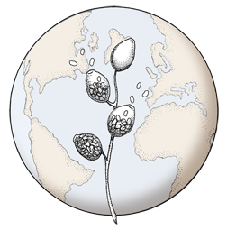

Paragynous antheridium attached to oogonium with oospore
Photo Gallery
Site will be retired 9/1/2026
This site is no longer being developed and will be retired on September 1, 2026. Please contact us if you have any questions or would like to provide support to continue the project.
|
P. tentaculata oospores and antheridia  |
P. cinnamomi hyphal swellings 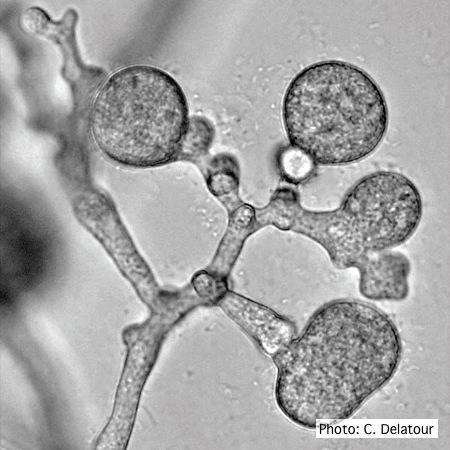 P. cinnamomi hyphal swellings (or thin walled chlamydospores) |
P. megakarya chlamydospore 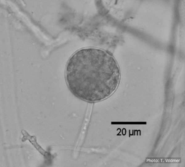
Terminal chlamydospore of P. megakarya
|
|
P. tentaculata microscopic characteristics 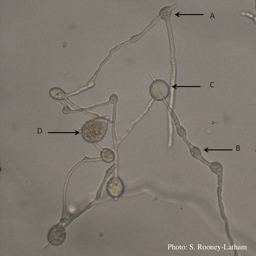 Hyphal swellings occuring at branching points of Mycelium (A), Intercalary hyphal swellings (B), Chlamydospore (C ), Sporangia (D) |
P. agathidicia oogonia 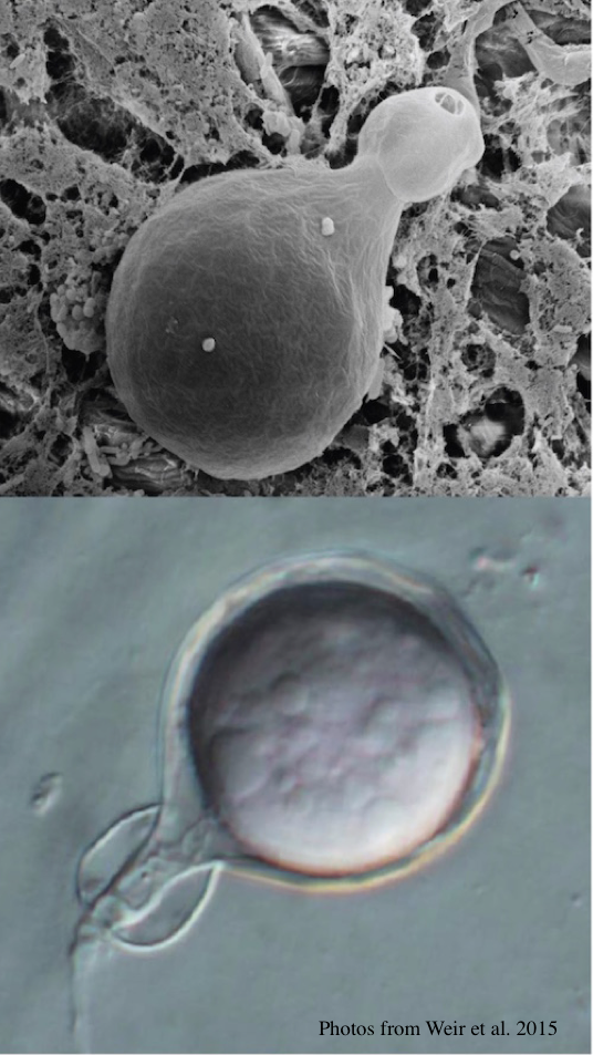 P. agathidicida oogonia with SEM (top) and light microscopy (bottom) |
P. arenaria sporangia  Globose papillate sporangia of Phytophthora arenaria on V8 agar flooded with soil extract. (Scale bar = 20 μm) |
|
P. pluvialis on Pinus radiata in New Zealand  Typical red needle cast symptoms along a twig. Lesions begin at the base of the needle which subsequently turns brown and is cast from the twig. |
P. siskiyouensis canker on Italian alder 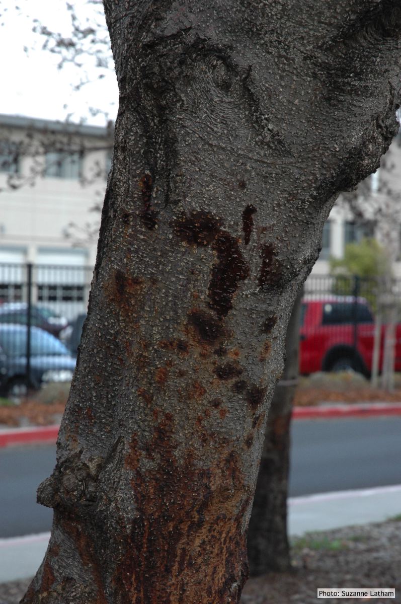 Phytophthora collar rot on Italian alder trees: an isolated bleeding canker on the trunk |
P. chlamydospora chlamydospore  Phytophthora chlamydospora chlamydospore in agar. Bar is 20µm. |
|
P. pinifolia colony morphology on PDA 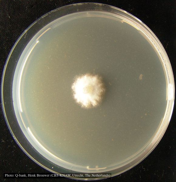 Colony pattern after 7 days on PDA at 24C, photo from Q-bank, used with permission. |
P. nicotianae sporangia  Noncaducous sporangium showing ovoid shape and papillate condition. (Fitopatol. bras. 2005) |
P. nicotianae chlamydospore 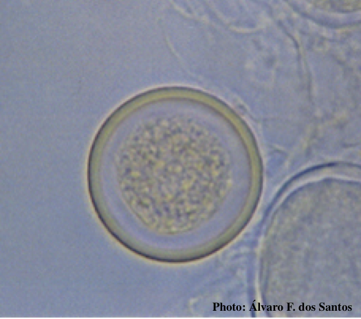 Globose chlamydospore (Fitopatol. bras. 2005) |