
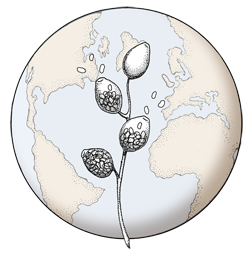

Broadly ovoid, papillate sporangia in water.
Photo Gallery
Site will be retired 9/1/2026
This site is no longer being developed and will be retired on September 1, 2026. Please contact us if you have any questions or would like to provide support to continue the project.
|
P. cactorum sporangia 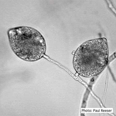 |
P. arenaria sporangia 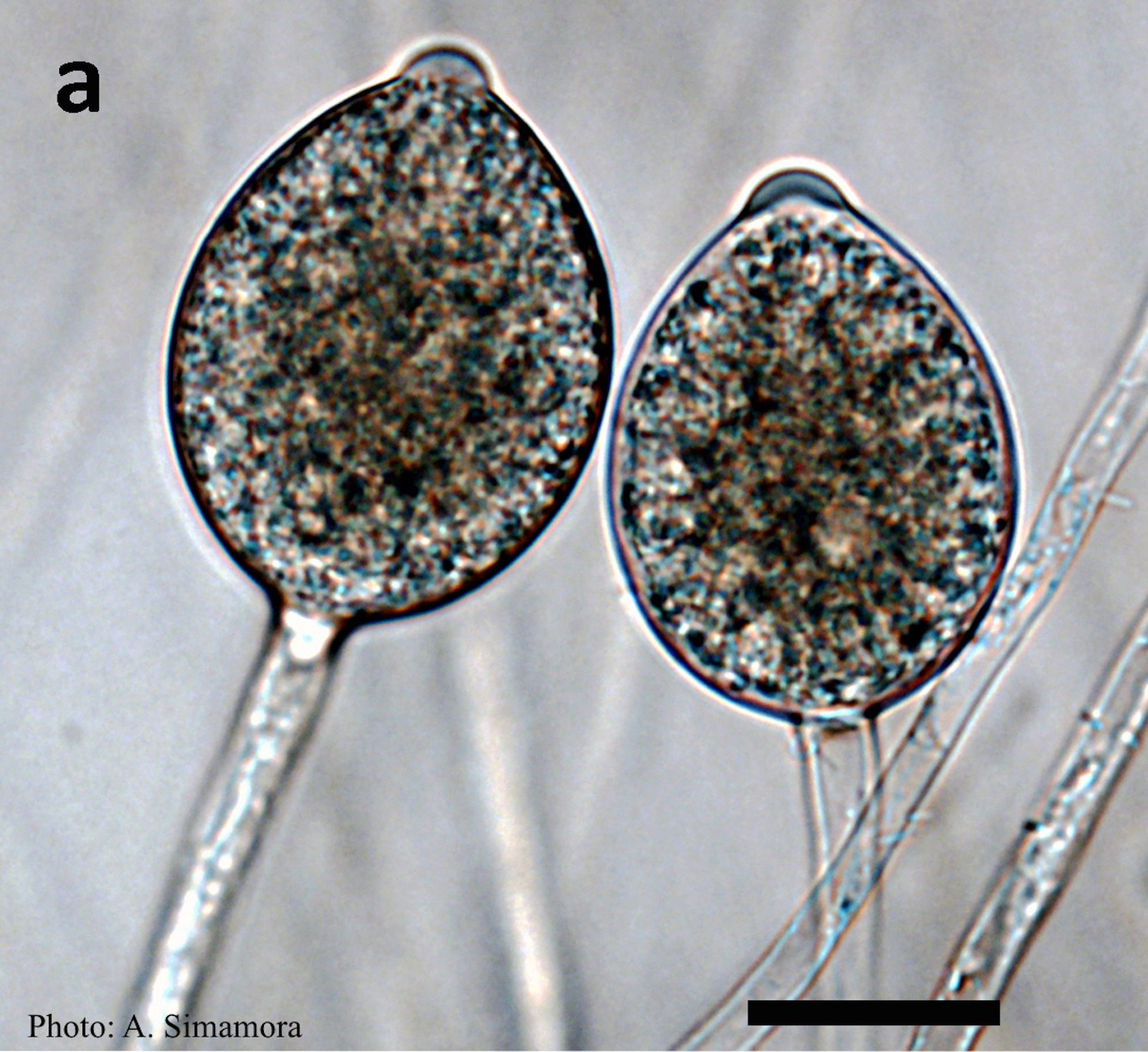 Typical ovoid papillate sporangia of Phytophthora arenaria on V8 agar flooded with soil extract |
Basal canker on Port Orford Cedar stump  |
|
P. arenaria sporangia  Globose papillate sporangia of Phytophthora arenaria on V8 agar flooded with soil extract. (Scale bar = 20 μm) |
P. frigida symptoms 3 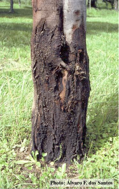 Black wattle bark with symptoms of gummosis |
P. tentaculata disease symptoms on California mugwort 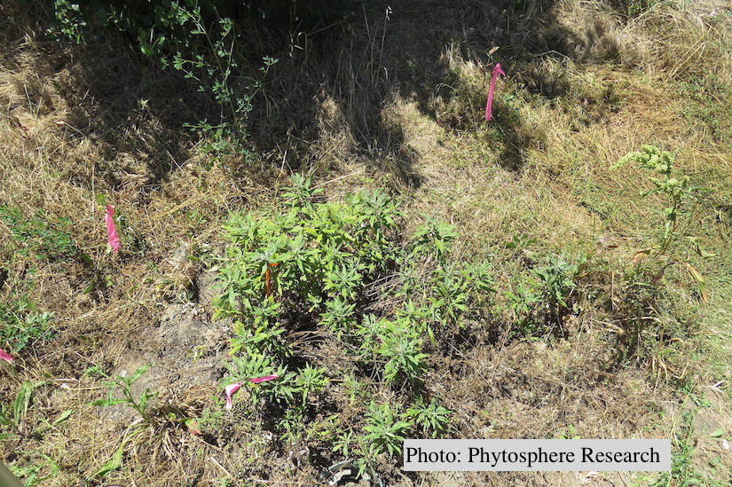 Outplanted California mugwort (Artemisia douglasiana) infected with P. tentaculata, 4.5 years after planting. Plant shows stunting and chlorosis. (P. cryptogea and P. lacustris were also baited from roots/soil of this plant). |
|
P. cambivora sporangium 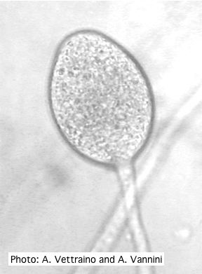 Ovoid non-papillate sporangia with well-rounded base and simple sporangiophore |
P. pinifolia sporangium 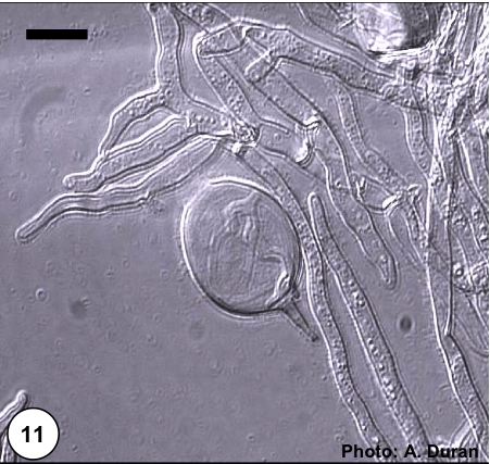 Non- papillate and caducous sporangia of Phytophthora pinifolia isolated from the infected P. radiata needles. |
P. katsurae oogonium 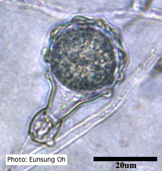 Warty protuberances on oogonium |
|
P. siskiyouensis bleeding canker  Bole lesions in the tissues under the bark of a bleeding canker: discoloration in the secondary phloem tissue |
P. ramorum colony morphology on CMA PARP 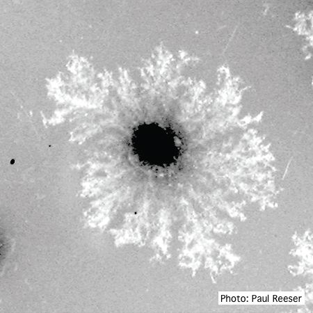 P. ramorum colony morphology on CMA PARP |
P. austrocedrae hyphal swellings 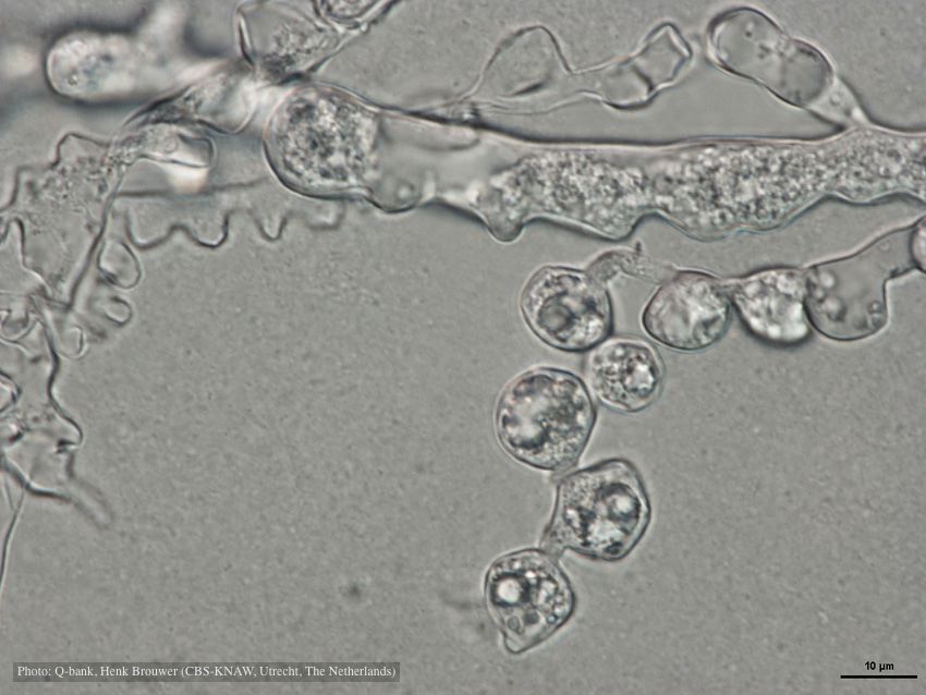 Hyphal swelling photo used with permission from Q-bank |