
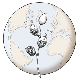

Bullate oogonium and and two-celled amphigynous antheridium
Photo Gallery
Site will be retired 9/1/2026
This site is no longer being developed and will be retired on September 1, 2026. Please contact us if you have any questions or would like to provide support to continue the project.
|
P. cambivora oogonium  |
P. cambivora colony morphology on PDA 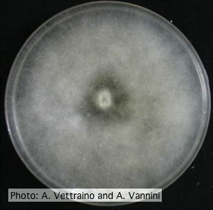 Uniform fluffy colony morphology at 14 days at 20°C on PDA |
P. cambivora sporangium 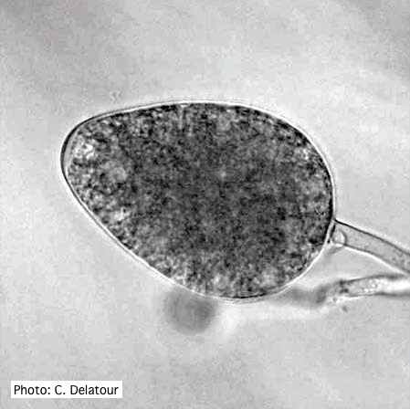 Ovoid non- papillate sporangia |
|
P. cambivora coralloid hyphae 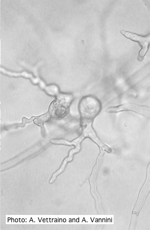 Coralloid hyphae with hyphal swelling-like structures |
P. cambivora symptoms 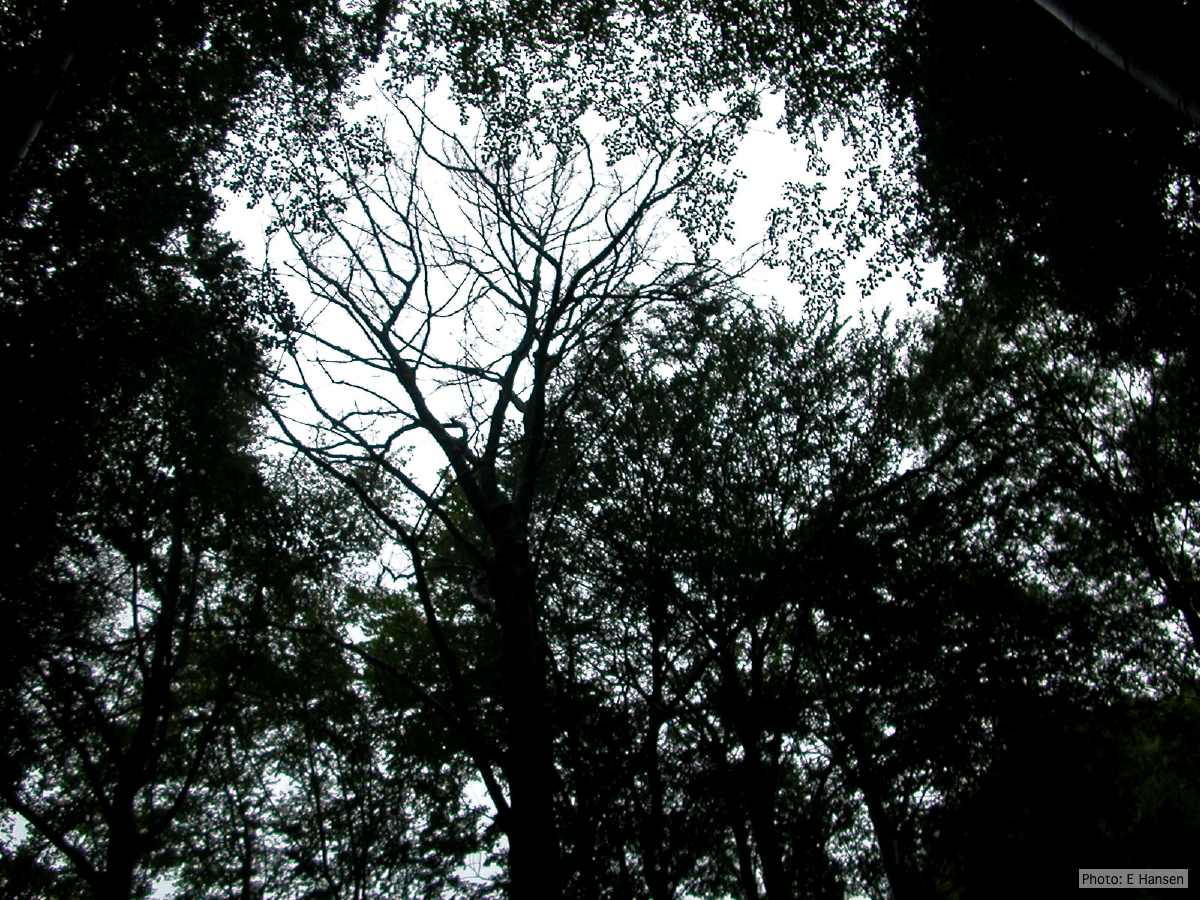 Dead beech in Germany |
P. cambivora sporangium 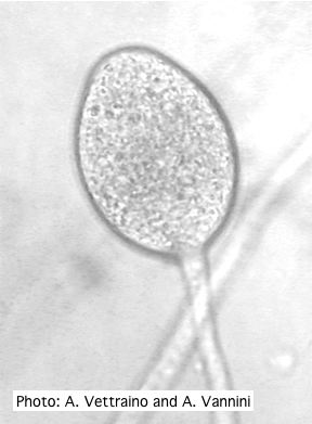 Ovoid non-papillate sporangia with well-rounded base and simple sporangiophore |
|
P. cambivora oogonium 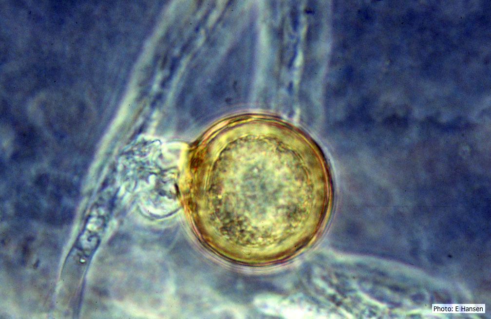 P. cambivora oogonium with antheridium |
P. cambivora disease symptoms 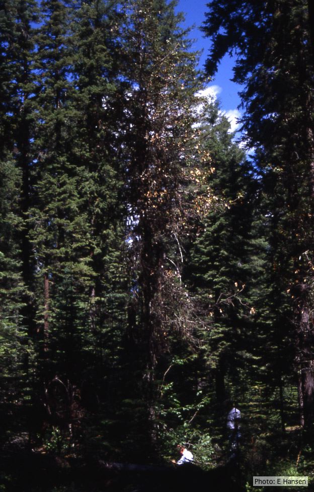 Dead and dying chinquapin infected with P. cambivora |
P. cambivora symptoms  P. cambivora on Fagus sylvatica bole |
|
P. cambivora sporangia 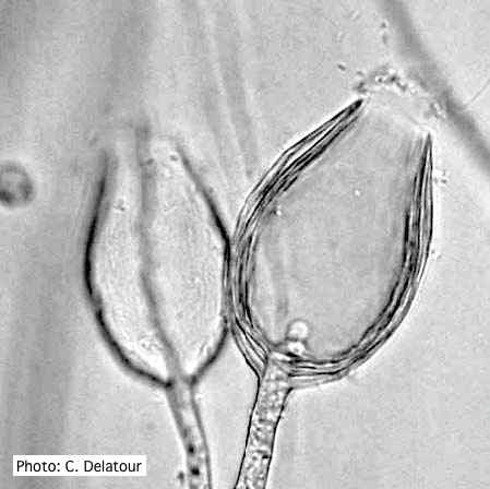 Empty sporangia of P. cambivora showing nested internal proliferation |
P. cambivora older hyphae  Old septate hyphae |
P. cambivora oogonium 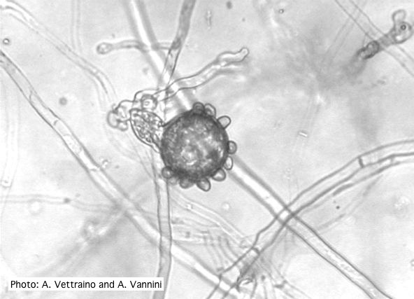 Bullate oogonium with amphyginous antheridium |