
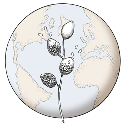

P. siskiyouensis sporangium with lateral semi-papilla and subterminal, sub-basal insertion in the sporangiophore
Photo Gallery
Site will be retired 9/1/2026
This site is no longer being developed and will be retired on September 1, 2026. Please contact us if you have any questions or would like to provide support to continue the project.
|
P. siskiyouensis sporangium  |
P. austrocedrae colony morphology on Tomato juice agar with B sitosterol 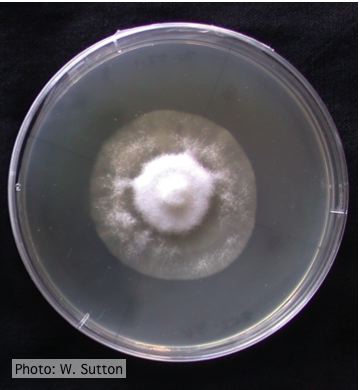 Colony morphology of P. austrocedrae at 16 ºC after 4 weeks on Tomato juice agar with B sitosterol |
P. cambivora tar spots 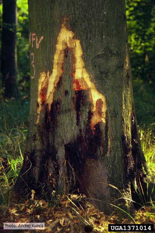 Tar spots on European beech (Fagus sylvatica) with bark removed. Lesse, Germany |
|
P. tentaculata oospores and antheridia  Paragynous antheridium attached to oogonium with oospore |
P. alni in riparian alder, Scotland 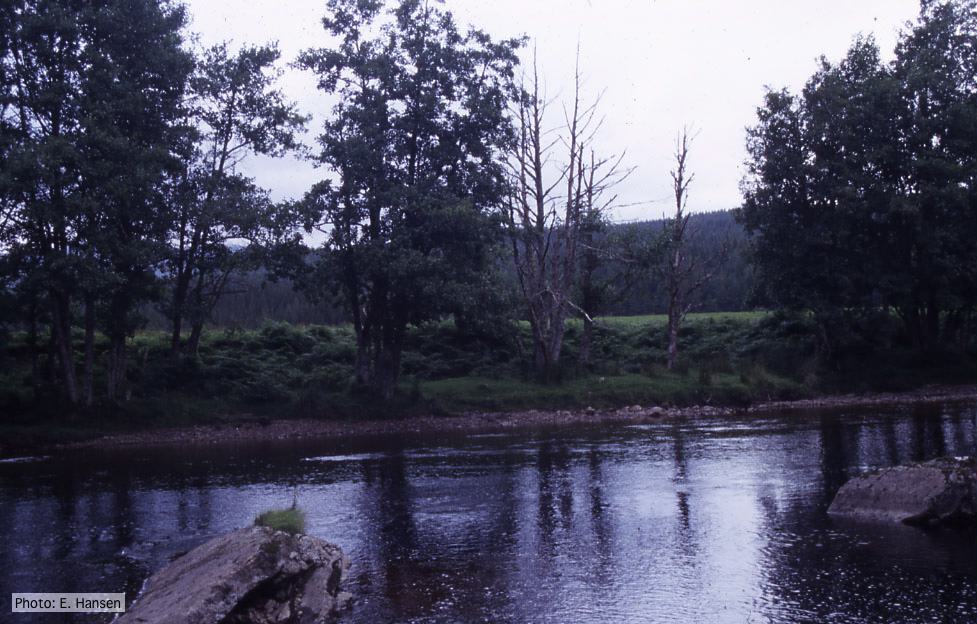 P. alni in riparian alder, Scotland |
P. pluvialis on Pinus radiata 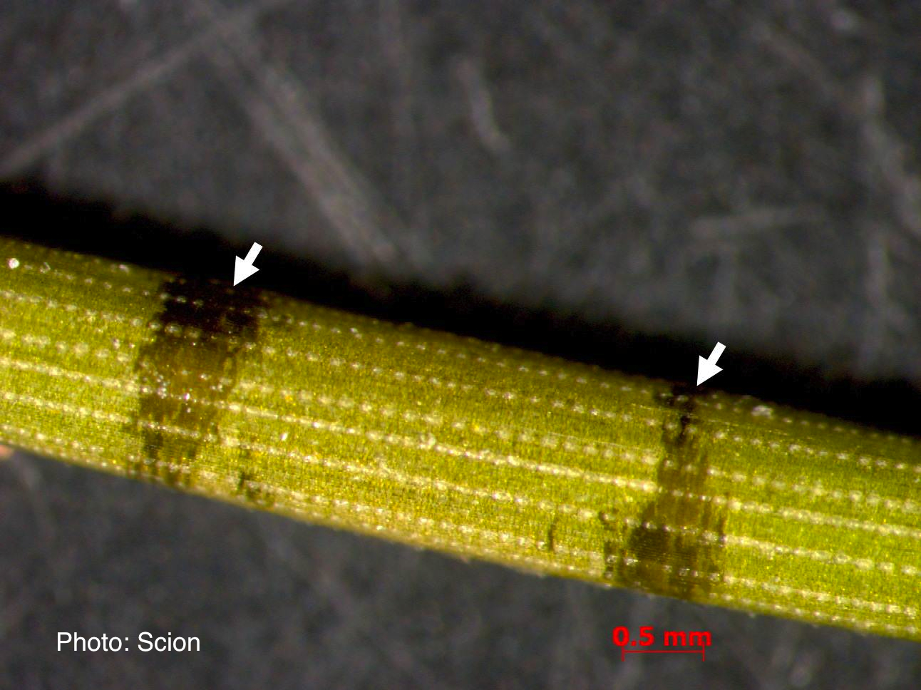 A Pinus radiata needle showing black resinous bands or marks consistent with the presence of red needle cast disease. |
|
P. palmivora colony morphology on V8 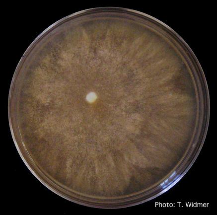 Growth of P. palmivora on V8 agar |
P. agathidicia growth on PDA 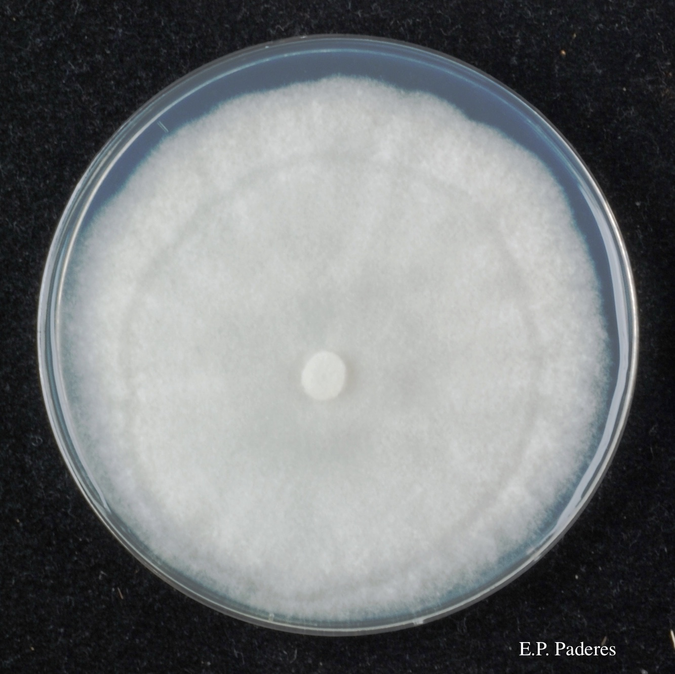 Colony morphology of ex-holotype ICMP 17027 after 10-days incubation at 20°C in the dark |
P. tentaculata on V-8 media 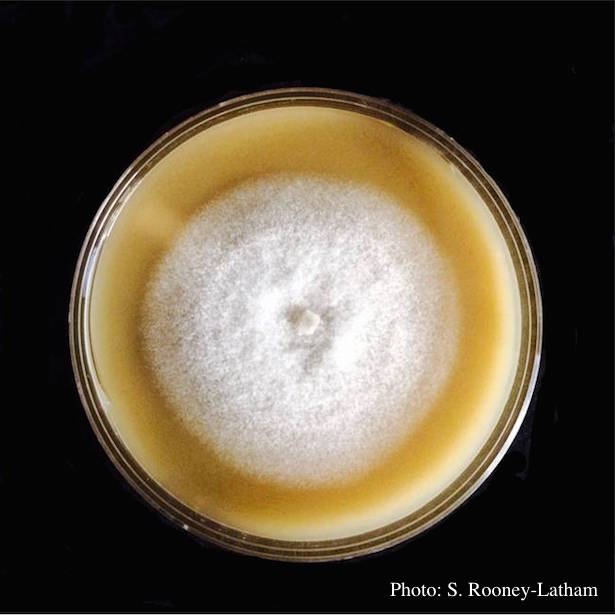 Culture of P. tentaculata on V-8 media |
|
P. megasperma oogonium 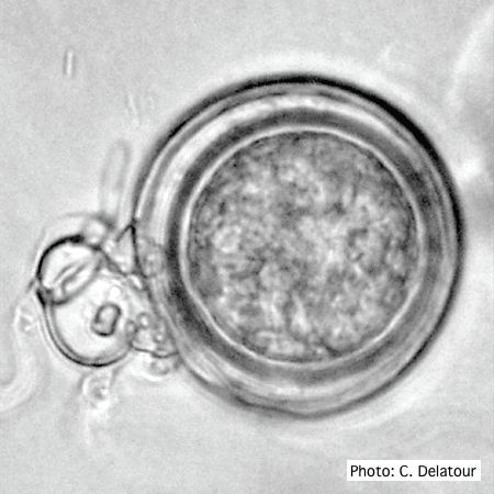 Oogonium with paragynous antheridia applied close to the ogonial stalk. |
P. cambivora oogonium 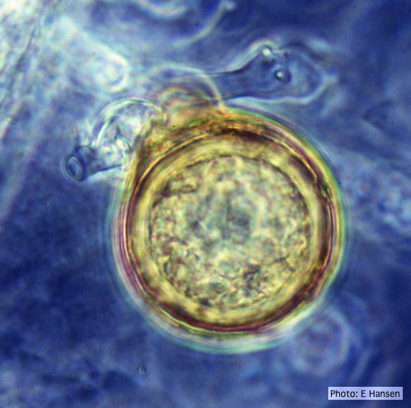 P. cambivora oogonium with antheridium |
P. megasperma colony morphology on PDA 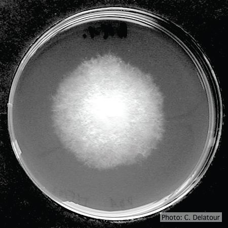 Colony morphology on PDA at 7 days |