

Close-up of margin area of bole lesions under the bark of a bleeding canker
Photo Gallery
Site will be retired 9/1/2026
This site is no longer being developed and will be retired on September 1, 2026. Please contact us if you have any questions or would like to provide support to continue the project.
|
P. siskiyouensis bleeding canker 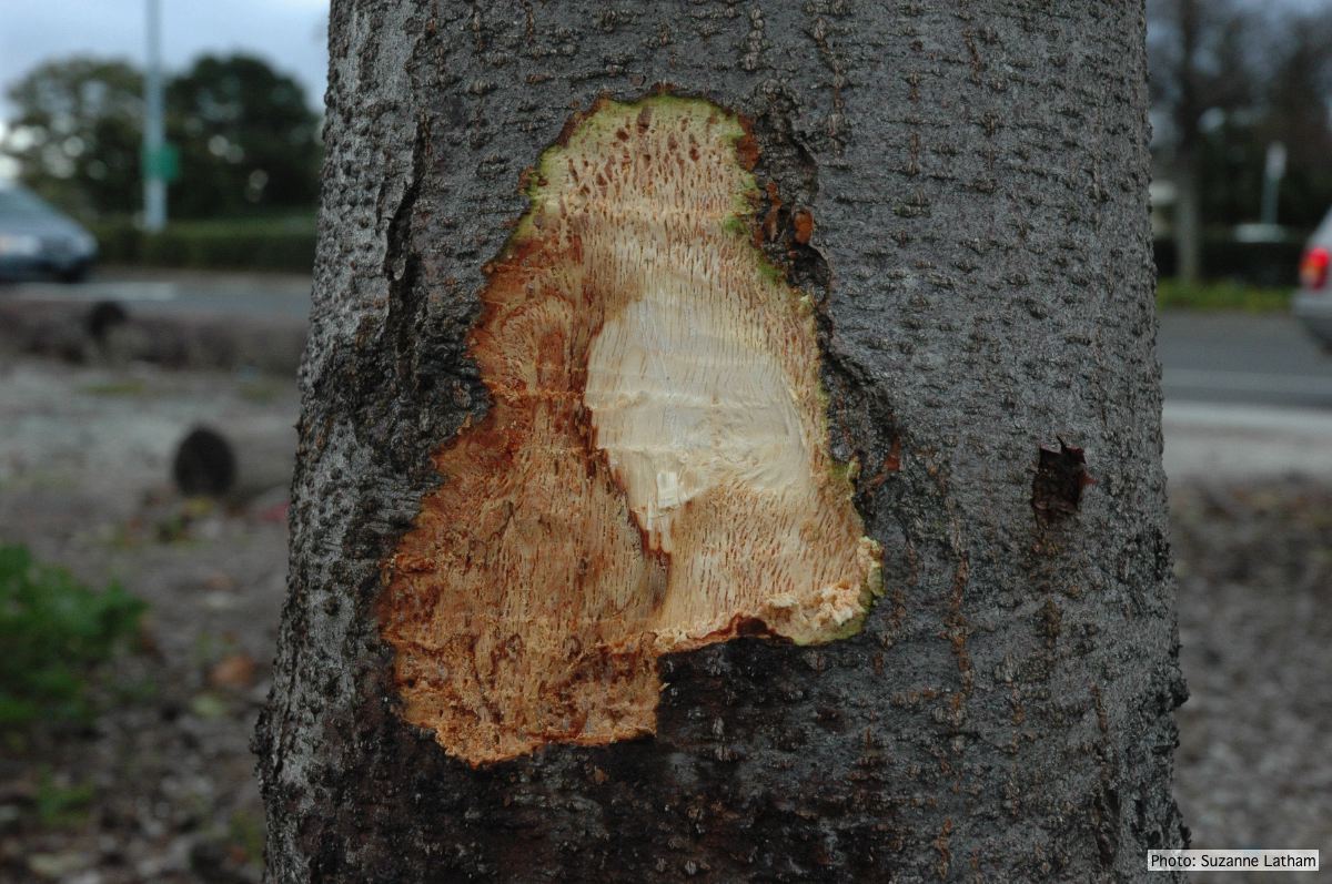 |
P. austrocedrae oogonium 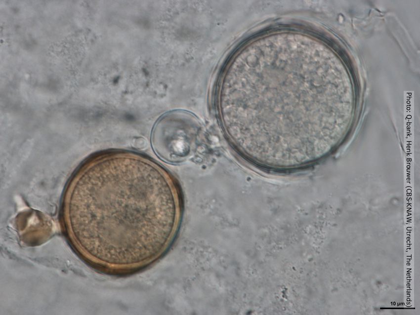 Oogonia with and without brown pigment, photo from Q-bank, used with permission |
P. cambivora oogonium 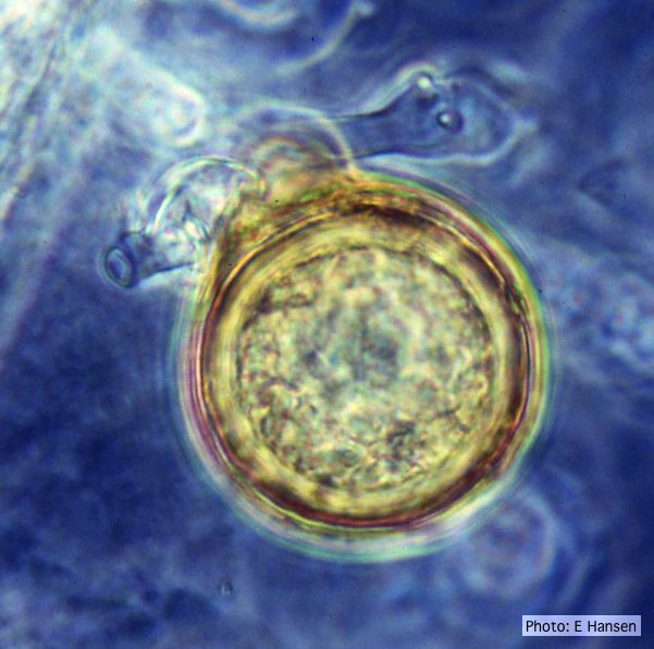 P. cambivora oogonium with antheridium |
|
P. ramorum sporangium  Deciduous sporangium, photo from Q-bank, used with permission |
P. nicotianae symptoms 2  Symptoms of gummosis on black wattle (Fitopatol. bras. 2005) |
P. pluvialis hyphal swellings 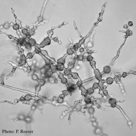 P. pluvialis hyphal swellings on agar |
|
P. agathidicida oospores  Oospores of P. agathidicida in the roots of kauri seedlings inoculated with P. agathidicida. The root has been cleared with potassium hydroxide and bleached with peroxide, before being stained with Trypan Blue |
P. pseudotsugae amphigynous oogonium 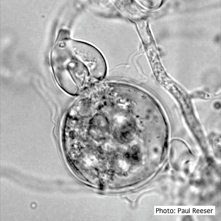 P. pseudotsugae oogonium with amphigynous antheridia |
P. cactorum oogonium 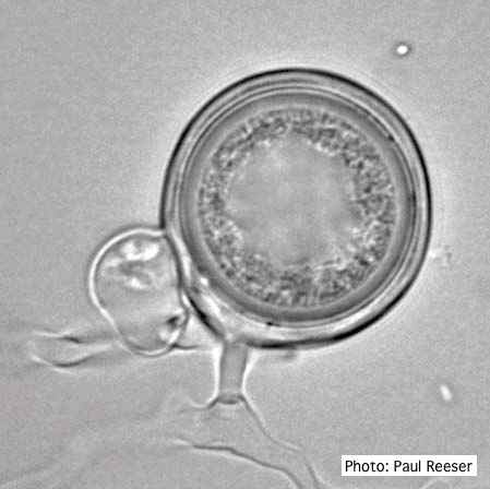 Oogonium with paragynous antheridia close to oogonial stalk. Oospores are slightly aplerotic. |
|
P. cambivora coralloid hyphae 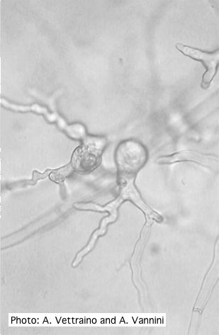 Coralloid hyphae with hyphal swelling-like structures |
P. kernoviae colony morphology on V8 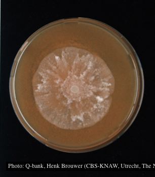 Colony morphology at 7 days at 18°C on V8, photo from Q-bank, used with permission. |
P. boehmeriae oogonium 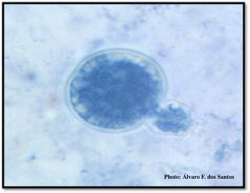 Oogonia and oospores with amphigynous antheridia |