
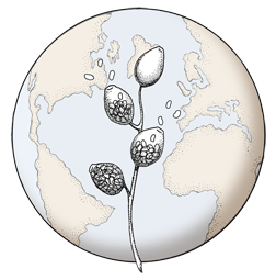

Growth of P. palmivora on corn meal agar
Photo Gallery
Site will be retired 9/1/2026
This site is no longer being developed and will be retired on September 1, 2026. Please contact us if you have any questions or would like to provide support to continue the project.
|
Growth of P. palmivora on CMA 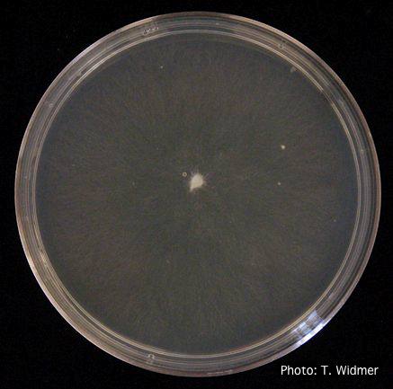 |
P. pinifolia colony morphology on PDA 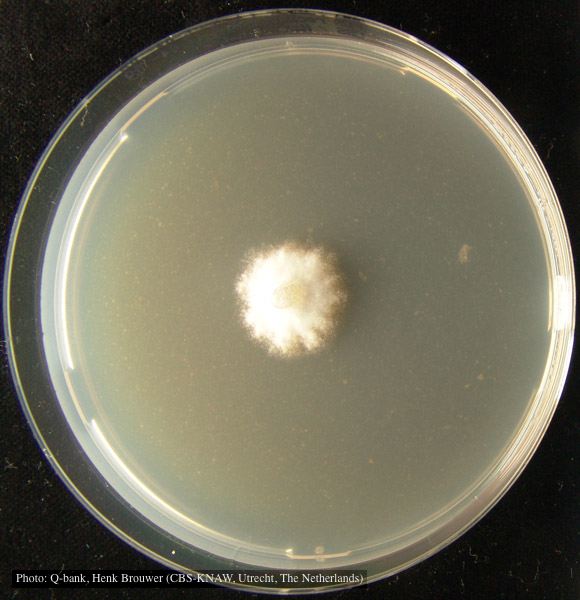 Colony pattern after 7 days on PDA at 24C, photo from Q-bank, used with permission. |
P. cambivora oogonium  Bullate oogonium and and two-celled amphigynous antheridium |
|
P. ramorum zoospores 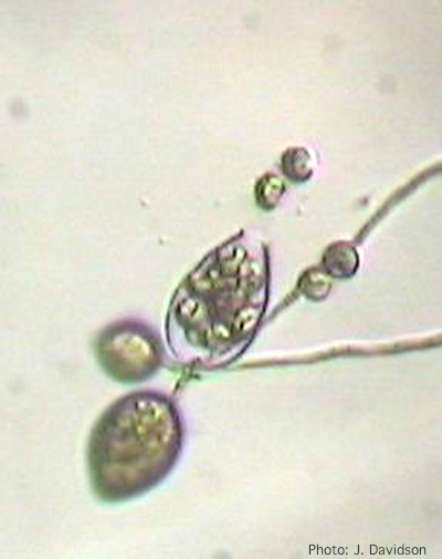 Sporangium of P. ramorum releasing zoospores |
P. frigida symptoms 3 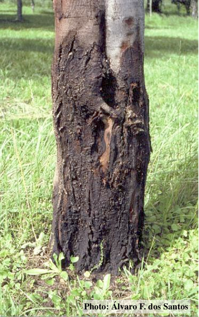 Black wattle bark with symptoms of gummosis |
P. pseudosyringae sporangia  Ovoid, semipapillate sporangia showing sympodial development of sporangiophore |
|
P. pluvialis on Pinus radiata needle  Clusters of sporangia emerge from stomata of an infected radiata pine needle. |
P. pseudosyringae colony morphology on PDA  Colony morphology on PDA at 14 days |
Comparative gametangial morphology of Phytophthora Clade 5 species 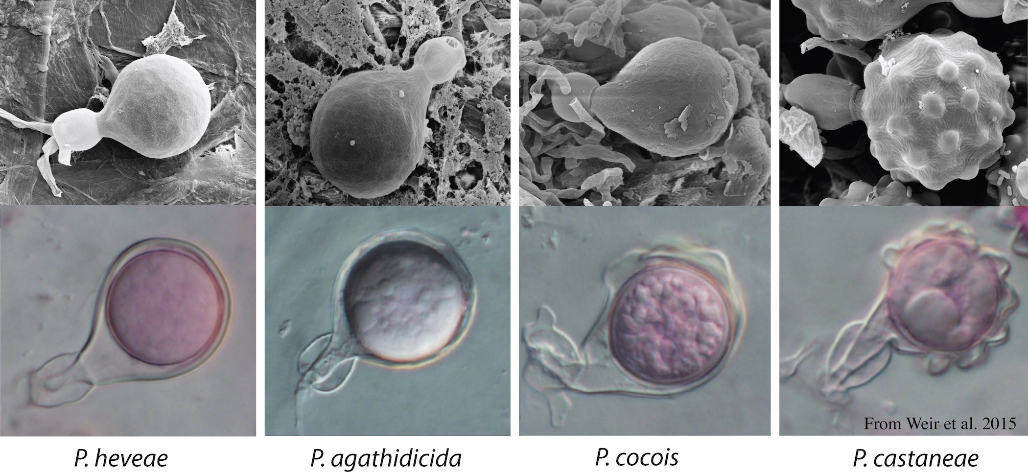 Comparative gametangial morphology of Phytophthora Clade 5 species, with SEM (top) and light microscopy (bottom). P. heveae has smooth walled oogonia with funnel-shaped, amphigynous antheridia. P. agathidicida has mildly stipulate oogonia with globose amphigynous antheridia. P.cocois has mildly bullate oogonia with reflexed amphigynous antheridia. P. castaneae has coarsely bullate oogonium with rugose protuberances and narrow amphigynous antheridia (Weir et al. 2015). |
|
P. pluvialis symptoms on Douglas-fir 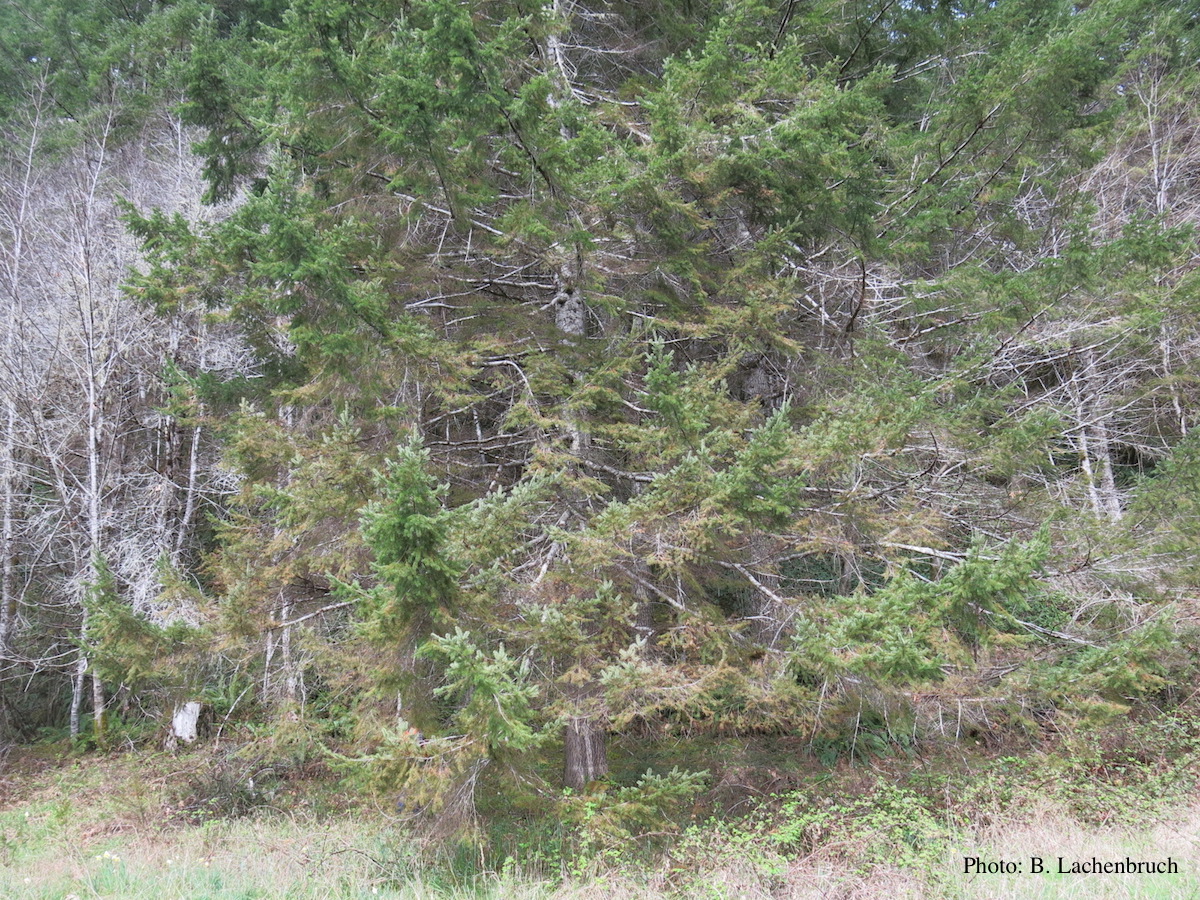 Red needle cast symptoms on Douglas-fir in western Oregon, 2015 |
P. megasperma colony morphology on V8  Colony morphology on V8 at 7 days |
P. chlamydospora hyphal swellings 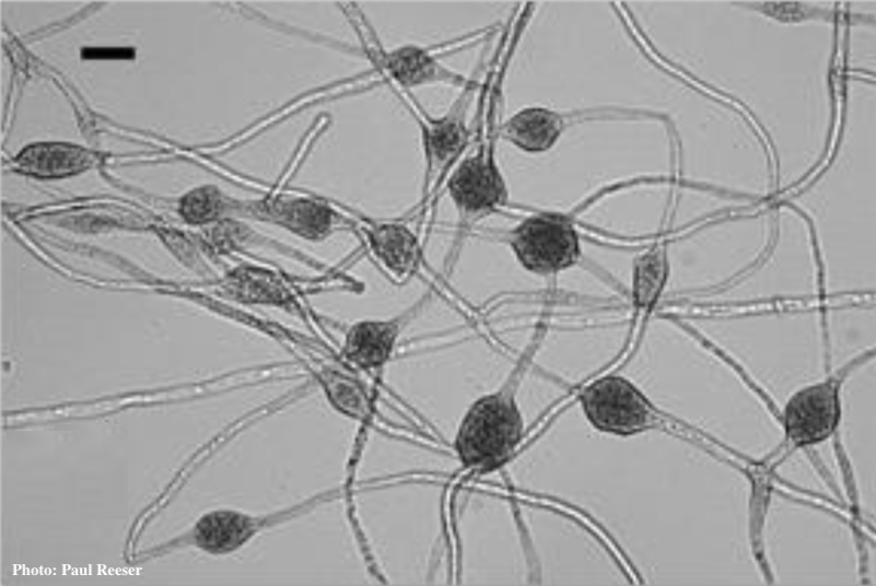 Phytophthora chlamydospora chlamydospore in agar. Bar is 20µm.
|