

Tufted appearance of new growth from surviving buds on Douglas-fir, one year after defoliation.
Photo Gallery
Site will be retired 9/1/2026
This site is no longer being developed and will be retired on September 1, 2026. Please contact us if you have any questions or would like to provide support to continue the project.
|
P. pluvialis - appearance of new growth 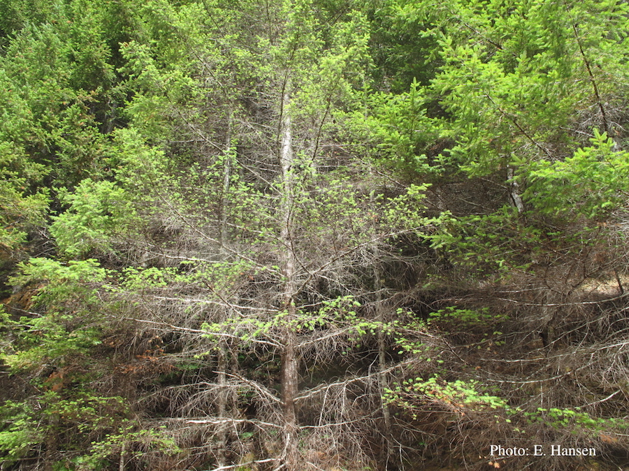 |
P. chlamydospora chlamydospore 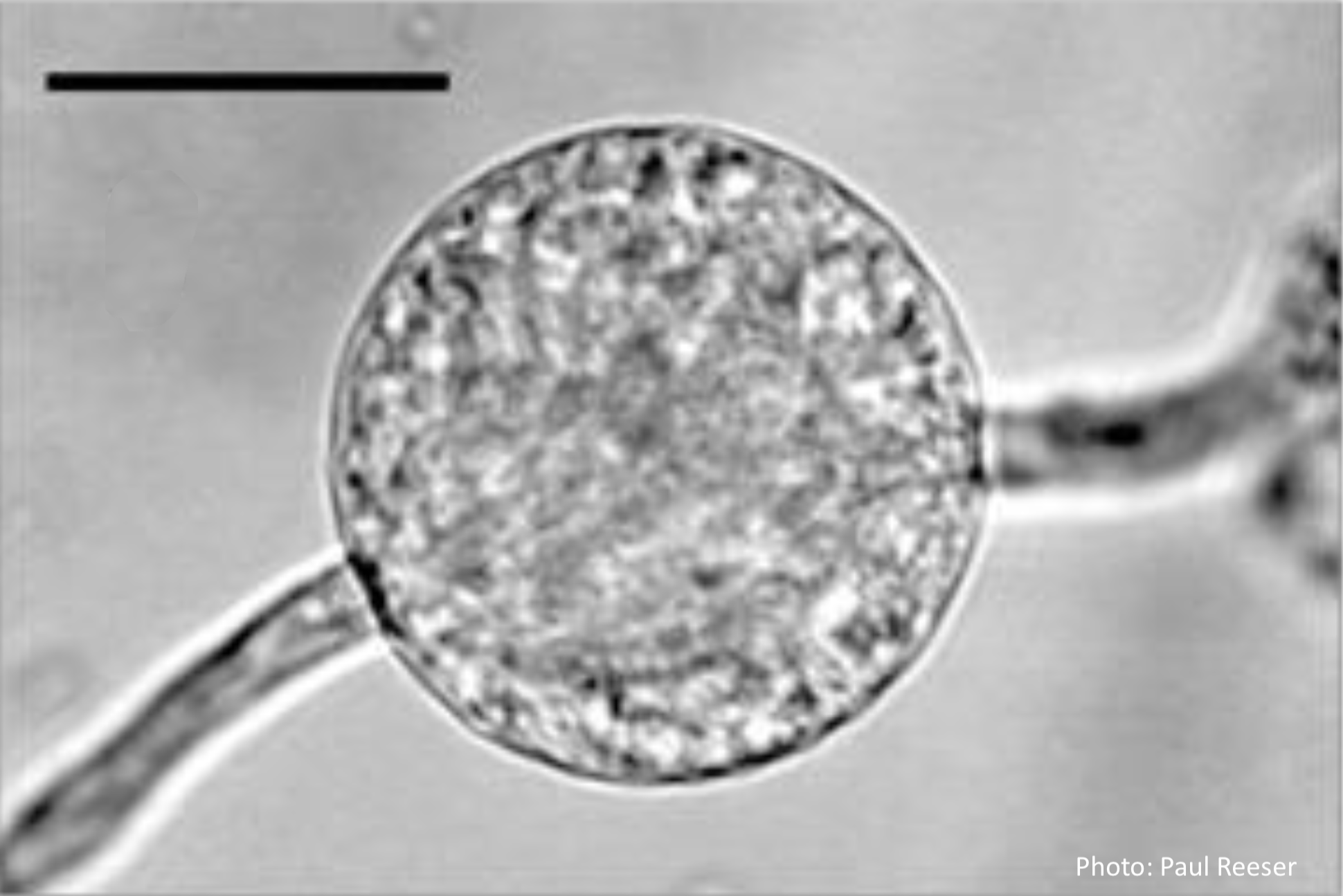 Phytophthora chlamydospora chlamydospore in agar. Bar is 20µm. |
P. agathidicia sporangia 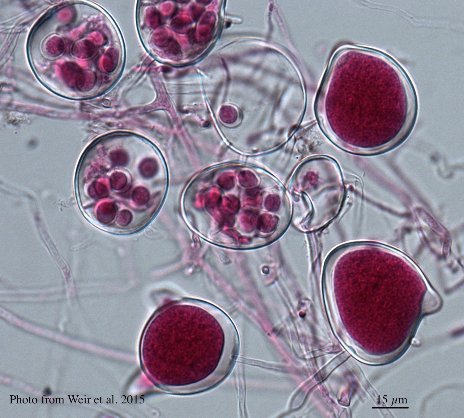 Differentiation of the cytoplasm within papillate sporangia into acid fuchsin stained zoospores |
|
P. katsurae growth morphology on β-CMA 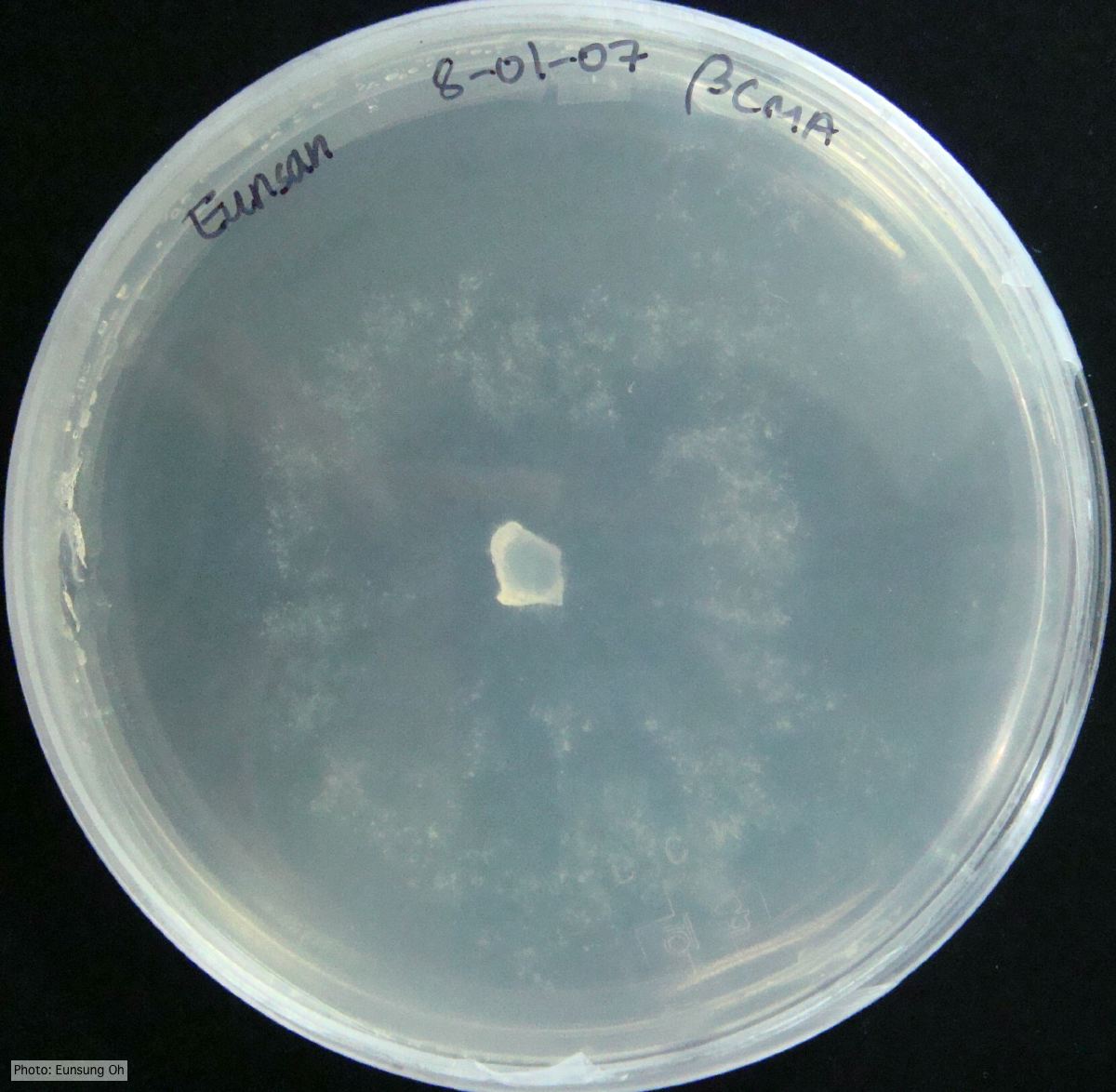 Growth morphology at 7 days on β-CMA |
P. chlamydospora chlamydospore  Phytophthora chlamydospora chlamydospore in agar. Bar is 20µm. |
P. lateralis sporangia 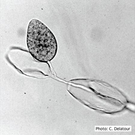 Sporangia of P. lateralis showing internal and external proliferation |
|
P. pluvialis symptoms on Douglas-fir 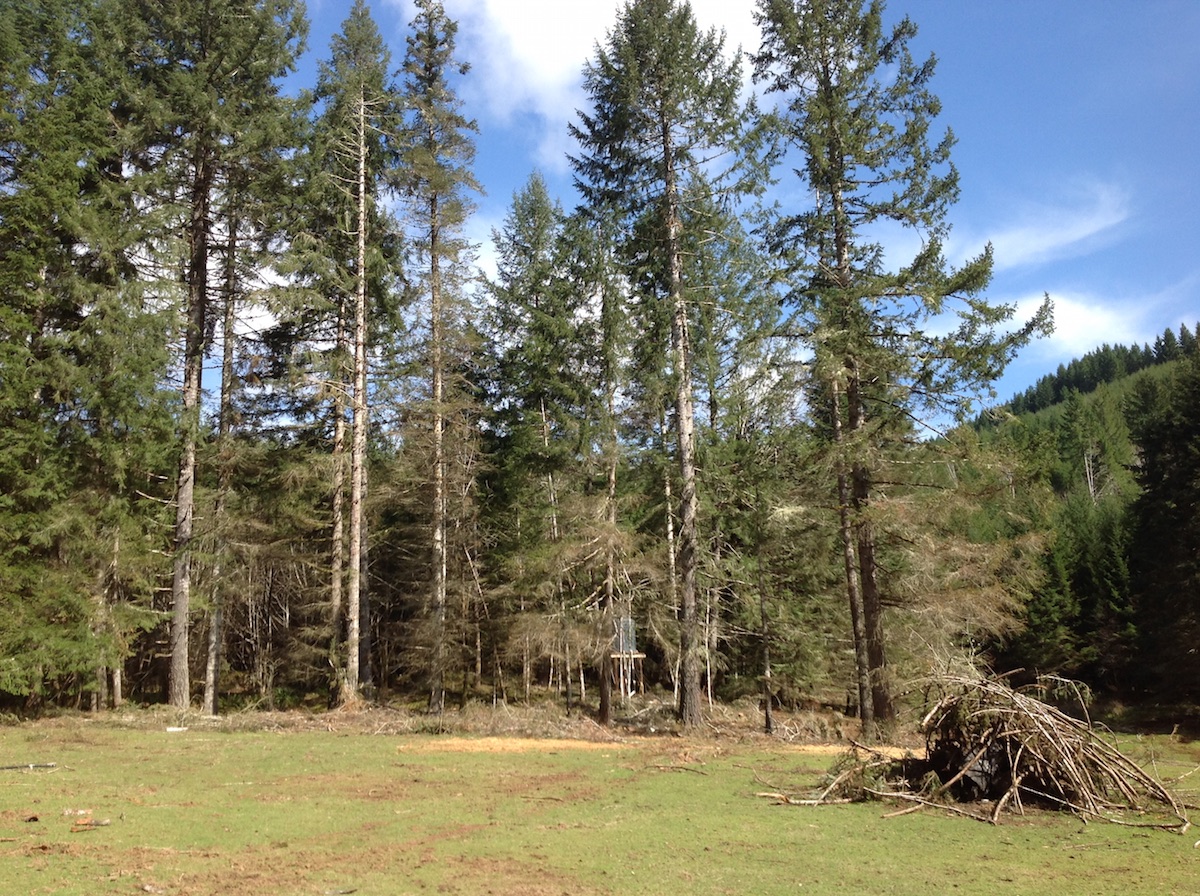 P. pluvialis symptoms of red needle cast on Douglas-fir, western Oregon 2015 |
P. siskiyouensis oogonium with amphigynous antheridium 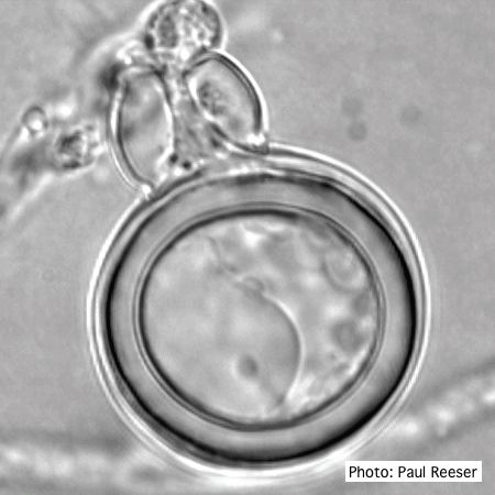 P. siskiyouensis oogonium with amphigynous antheridium |
P. siskiyouensis bleeding canker  Bole lesions in the tissues under the bark of a bleeding canker: discoloration in the secondary phloem tissue |
|
Growth of P. arenaria on half-strength PDA 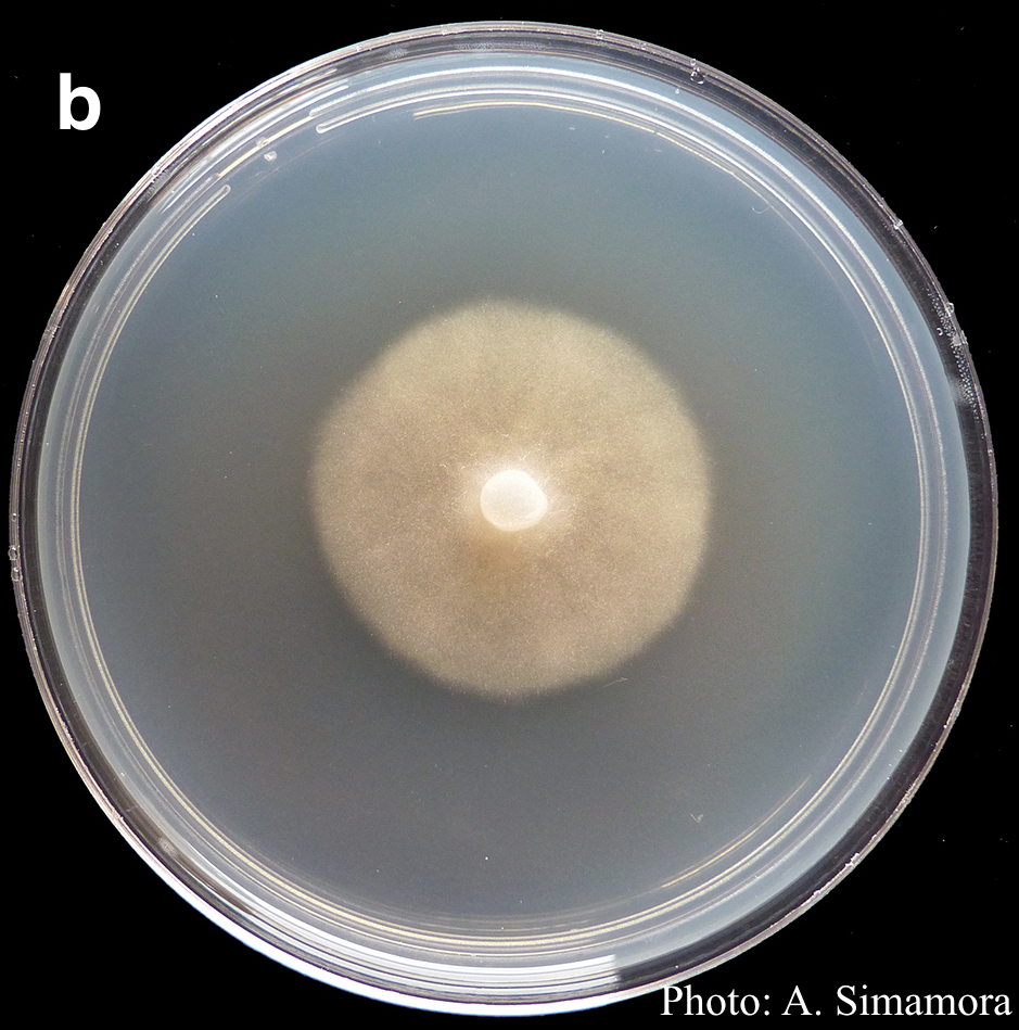 Colony morphology of Phytophthora arenaria after 7 days at 20°C on half-strength PDA |
P. pluvialis symptoms on Douglas-fir 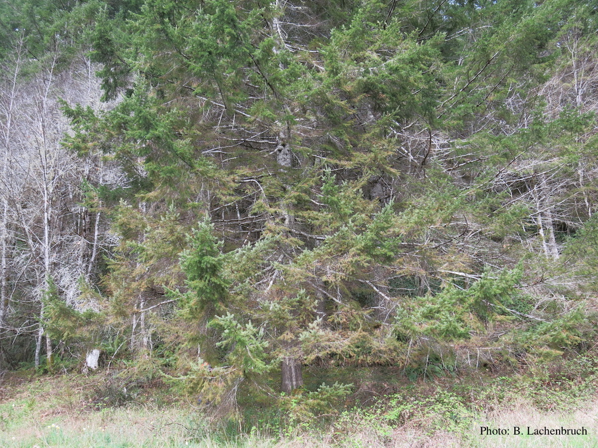 Red needle cast symptoms on Douglas-fir in western Oregon, 2015 |
P. cactorum bleeding canker 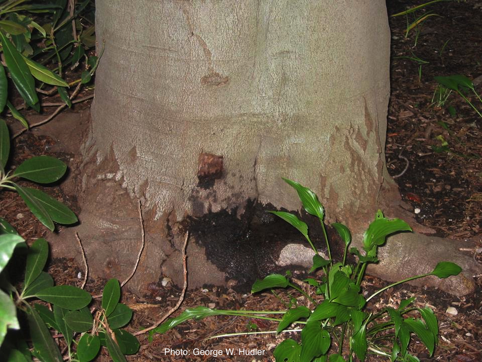 Bleeding canker on European beech (Fagus sylvatica) |