

Jogger running over a plastic-reinforced, foam-mat containing a 2% solution of Trigene™ Advance (quaternary ammonium compound) as part of a cross-country event, in the Waitakere Regional Park
Photo Gallery
Site will be retired 9/1/2026
This site is no longer being developed and will be retired on September 1, 2026. Please contact us if you have any questions or would like to provide support to continue the project.
|
Mat to control spread of P. agathidicida 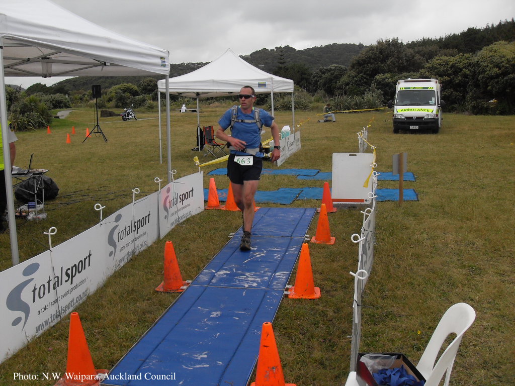 |
Boot wash to station to control spread of P. agathidicida 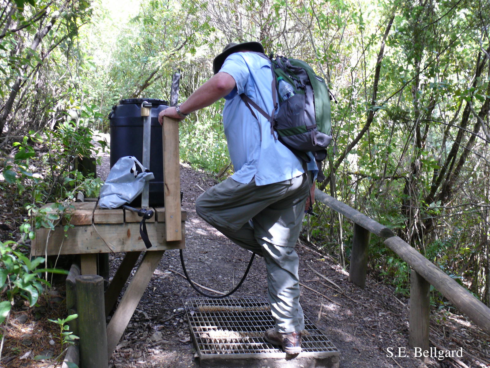 Use of hypochlorite solution applied through a “livestock drench-gun”, integrated with a soil grate to allow potentially contaminated soil to be collected |
P. agathidicia sporangia 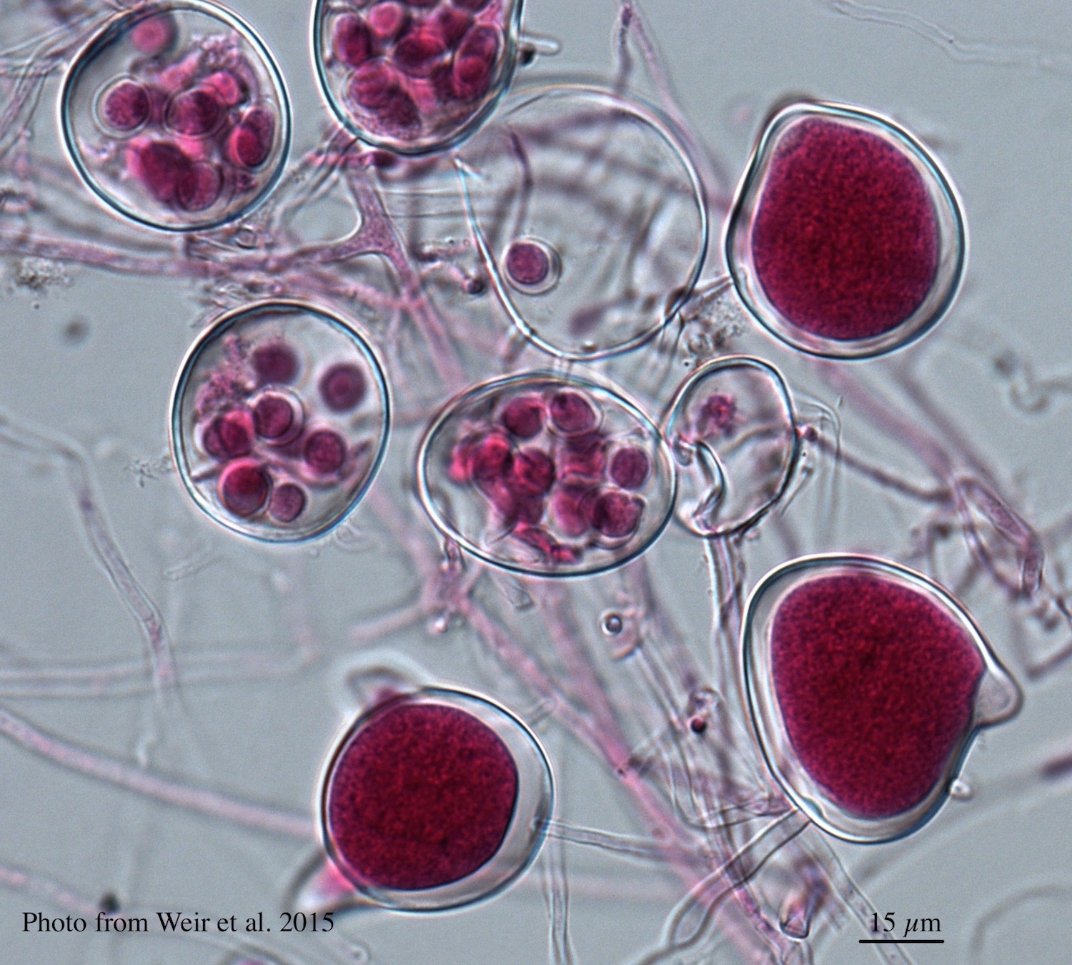 Differentiation of the cytoplasm within papillate sporangia into acid fuchsin stained zoospores |
|
P. agathidicida lesion on kauri tree 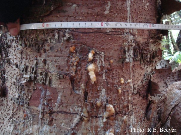 Close up of gum oozing out of lower trunk lesions of a young kauri tree at Maungaroa Ridge, Piha region of Waitakere Regional Park |
P. agathidicia oogonia 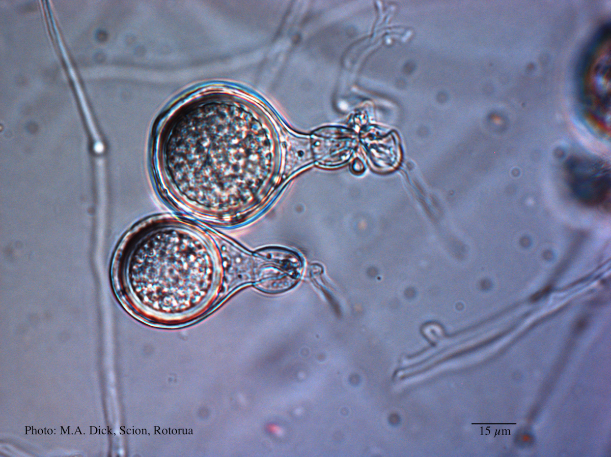 Light micrograph of P. agathidicida oospore (Scale bar equals 15 µm) |
P. agathidicida lesion on kauri tree 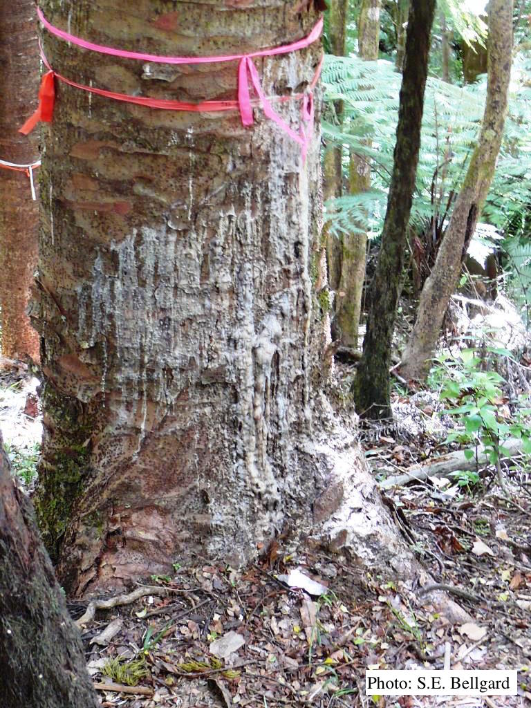 Advancing triangular lesion extending up the trunk of an 80 cm DBH kauri tree in the Huia Dam Site along Twin Peaks Track, Waitakere Regional Park |
|
P. agathidicida lesion on kauri tree 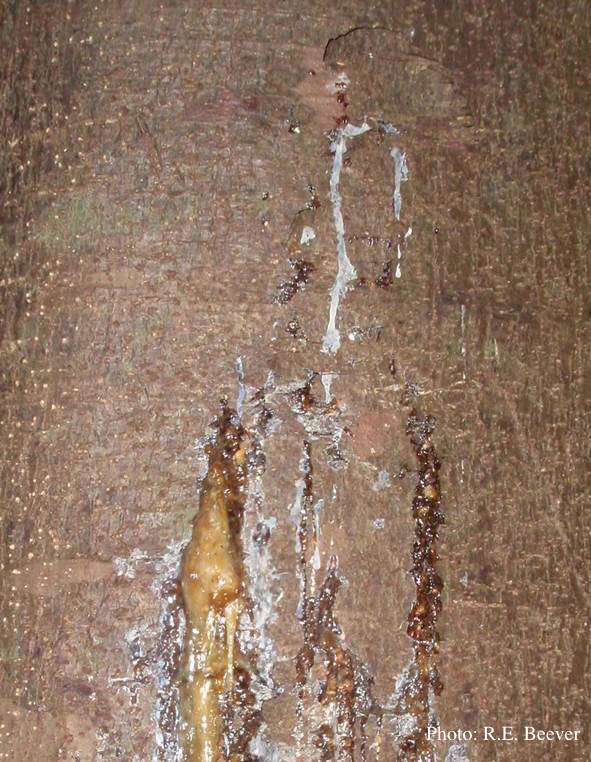 Gum oozing out of longitudinal lesion |