

Comparative gametangial morphology of Phytophthora Clade 5 species, with SEM (top) and light microscopy (bottom). P. heveae has smooth walled oogonia with funnel-shaped, amphigynous antheridia. P. agathidicida has mildly stipulate oogonia with globose amphigynous antheridia. P.cocois has mildly bullate oogonia with reflexed amphigynous antheridia. P. castaneae has coarsely bullate oogonium with rugose protuberances and narrow amphigynous antheridia (Weir et al. 2015).
Photo Gallery
Site will be retired 9/1/2026
This site is no longer being developed and will be retired on September 1, 2026. Please contact us if you have any questions or would like to provide support to continue the project.
|
Comparative gametangial morphology of Phytophthora Clade 5 species  |
Boot wash to station to control spread of P. agathidicida 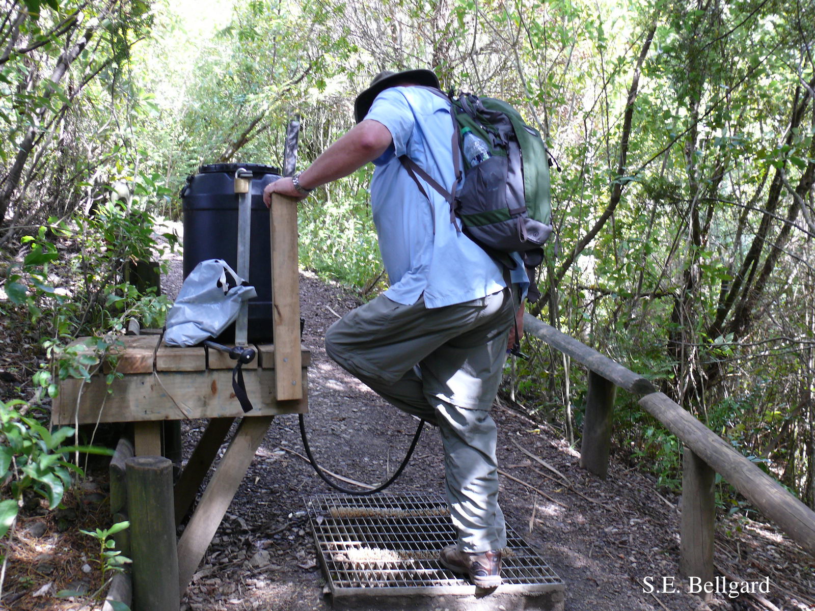 Use of hypochlorite solution applied through a “livestock drench-gun”, integrated with a soil grate to allow potentially contaminated soil to be collected |
Mat to control spread of P. agathidicida  Jogger running over a plastic-reinforced, foam-mat containing a 2% solution of Trigene™ Advance (quaternary ammonium compound) as part of a cross-country event, in the Waitakere Regional Park |
|
P. agathidicida oospores  Oospores of P. agathidicida in the roots of kauri seedlings inoculated with P. agathidicida. The root has been cleared with potassium hydroxide and bleached with peroxide, before being stained with Trypan Blue |
P. agathidicia sporangia 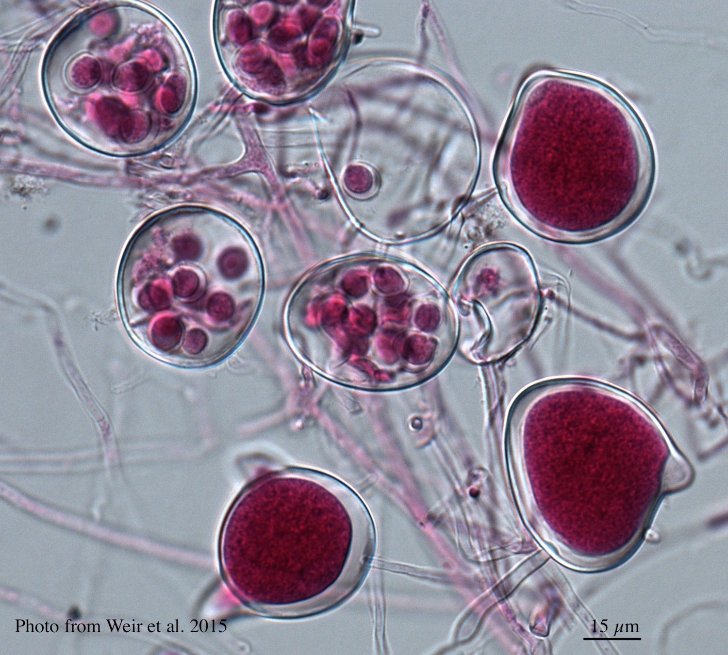 Differentiation of the cytoplasm within papillate sporangia into acid fuchsin stained zoospores |
P. agathidicia growth on PDA 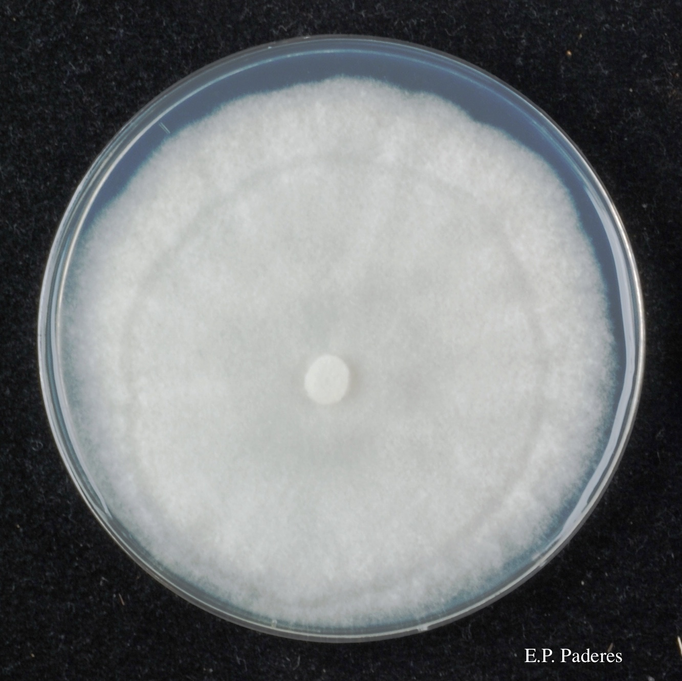 Colony morphology of ex-holotype ICMP 17027 after 10-days incubation at 20°C in the dark |
|
P. agathidicida lesion on kauri tree 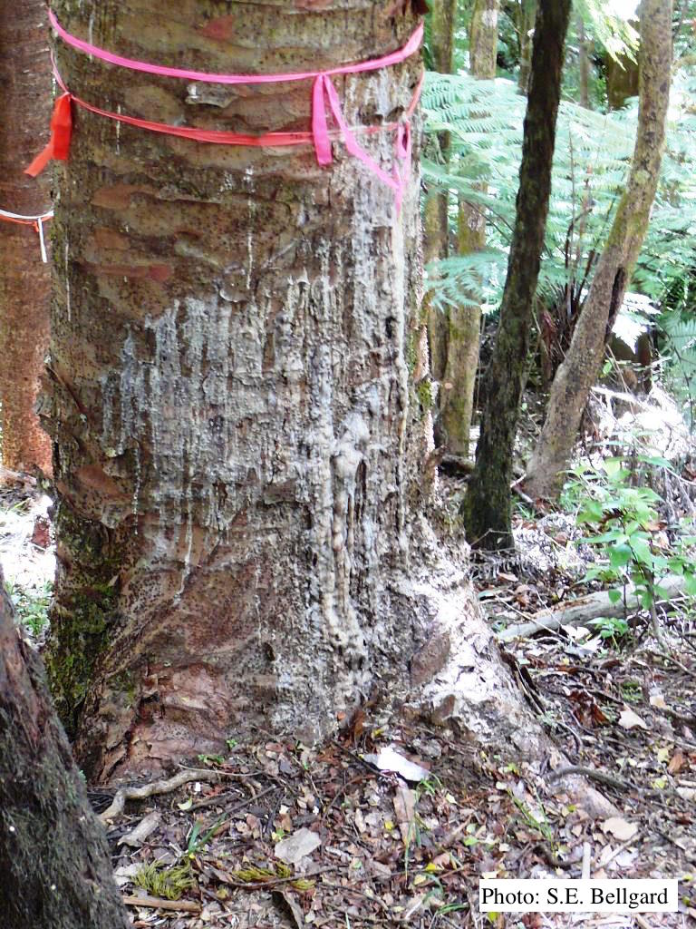 Advancing triangular lesion extending up the trunk of an 80 cm DBH kauri tree in the Huia Dam Site along Twin Peaks Track, Waitakere Regional Park |
P. agathidicida lesion on kauri tree 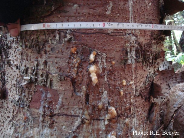 Close up of gum oozing out of lower trunk lesions of a young kauri tree at Maungaroa Ridge, Piha region of Waitakere Regional Park |
P. agathadicida disease symptom 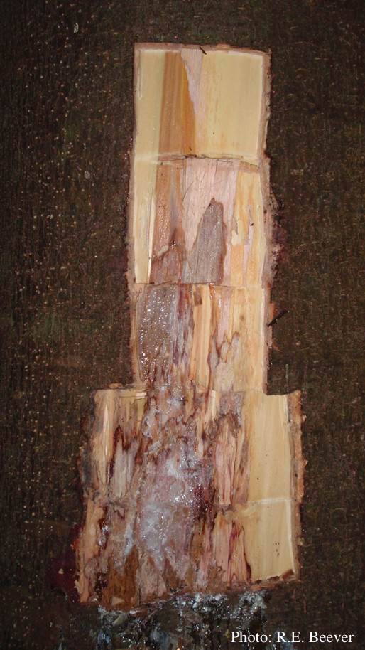 Excavated lesion, with outer bark removed showing extent of disease-front |
|
Tāne Mahuta “Lord of the Forest” kauri tree 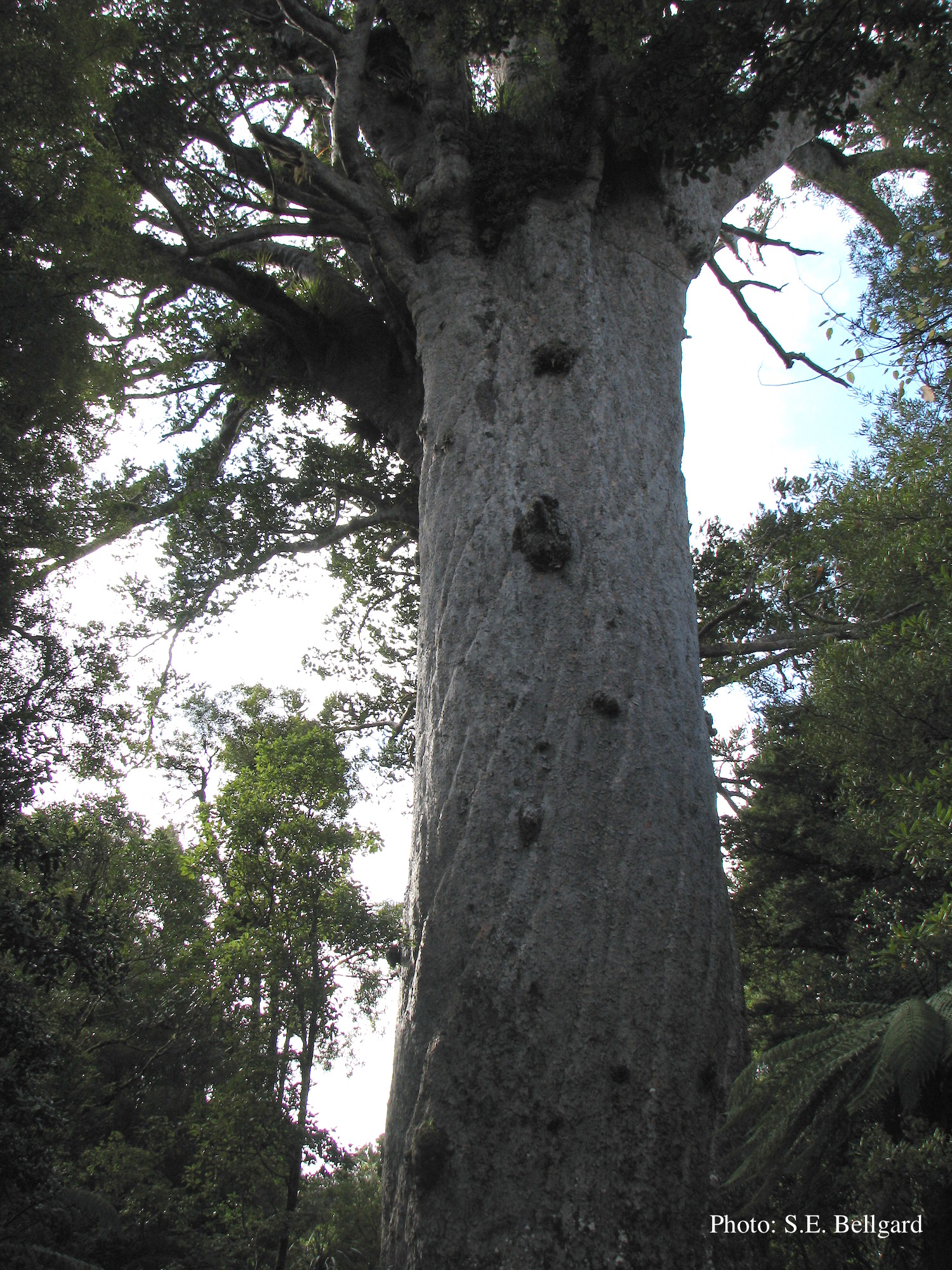 Tāne Mahuta “Lord of the Forest” is a giant kauri tree (approximately 47 metres in height) in the Waipoua Forest of Northland Region, New Zealand. Its age is unknown but is estimated to be between 1,250 and 2,500 years |
P. agathidicia disease symptoms on kauri 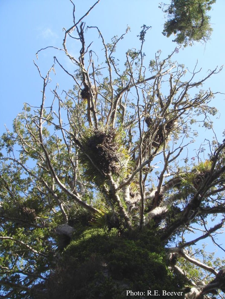 Crown decline of mature kauri, with branchlets with little or no leaves |
P. agathidicida growth on MEA  Diffuse, non-patterned, colony morphology of ICMP 16471 (the original “Gadgil isolate”) after 10-days incubation at 20°C in the dark |