

P. cactorum sporangia
Photo Gallery
Site will be retired 9/1/2026
This site is no longer being developed and will be retired on September 1, 2026. Please contact us if you have any questions or would like to provide support to continue the project.
|
P. cactorum sporangia  |
P. katsurae disease symptoms 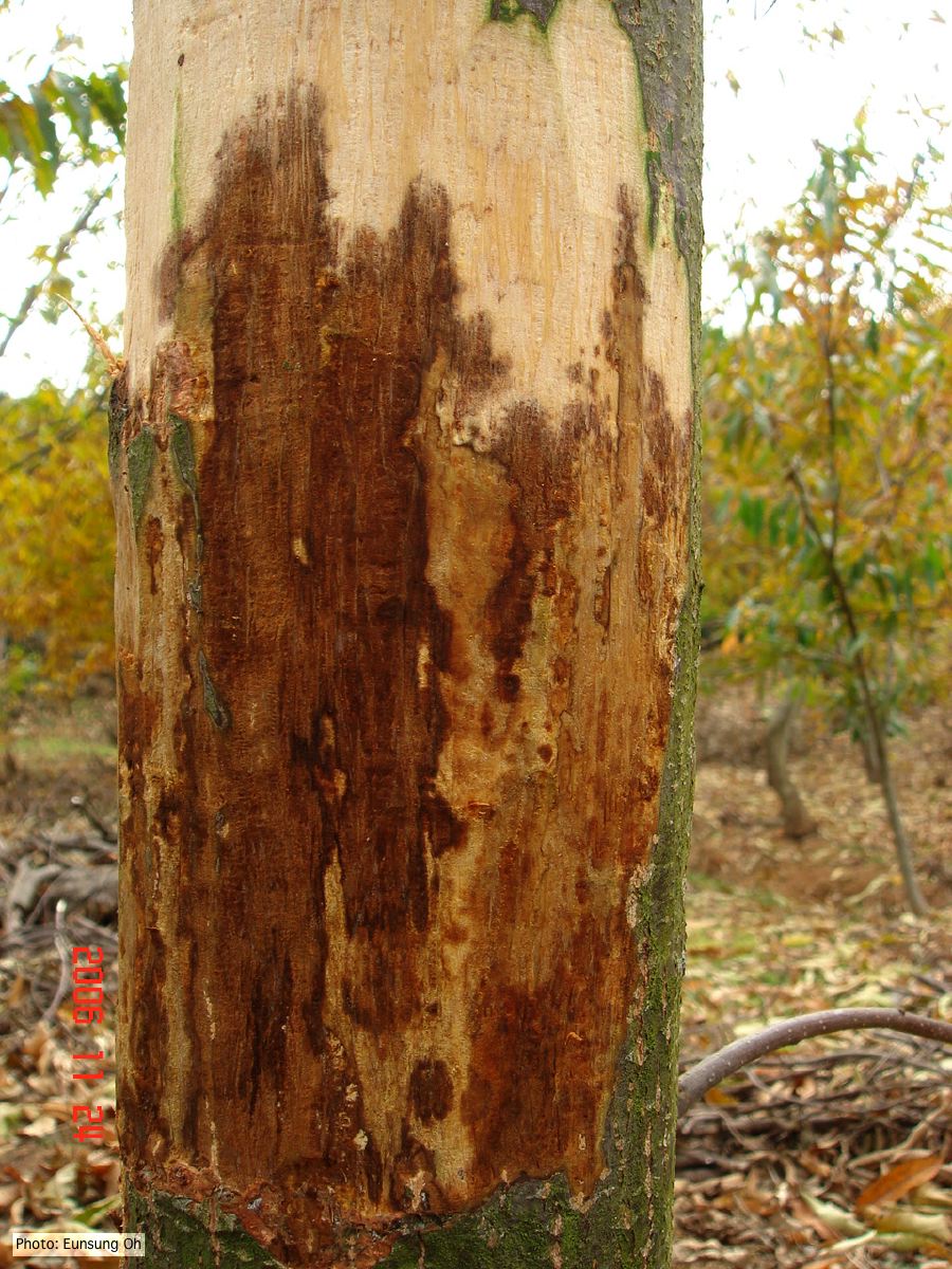 Infected chestnut (Castanea) with girdling canker |
Growth of P. arenaria on CA  Colony morphology of Phytophthora arenaria after 7 days at 20°C on carrot agar |
|
Comparative gametangial morphology of Phytophthora Clade 5 species  Comparative gametangial morphology of Phytophthora Clade 5 species, with SEM (top) and light microscopy (bottom). P. heveae has smooth walled oogonia with funnel-shaped, amphigynous antheridia. P. agathidicida has mildly stipulate oogonia with globose amphigynous antheridia. P.cocois has mildly bullate oogonia with reflexed amphigynous antheridia. P. castaneae has coarsely bullate oogonium with rugose protuberances and narrow amphigynous antheridia (Weir et al. 2015). |
P. cinnamomi on Banksia 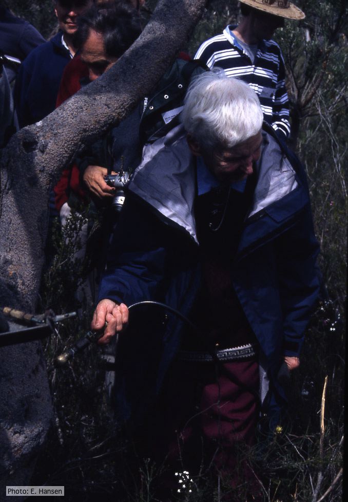 Gretna Weste injecting Banksia with phosphonate |
P. cambivora colony morphology on PDA 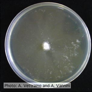 Appressed colony morphology at 14 days at 20°C on PDA |
|
Phytophthora taxon Agathis bole canker  Canker on a Kauri tree, New Zealand |
P. cinnamomi hyphal swellings 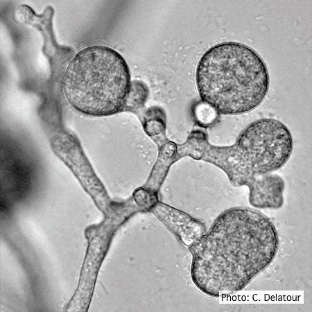 P. cinnamomi hyphal swellings (or thin walled chlamydospores) |
P. agathadicida disease symptom 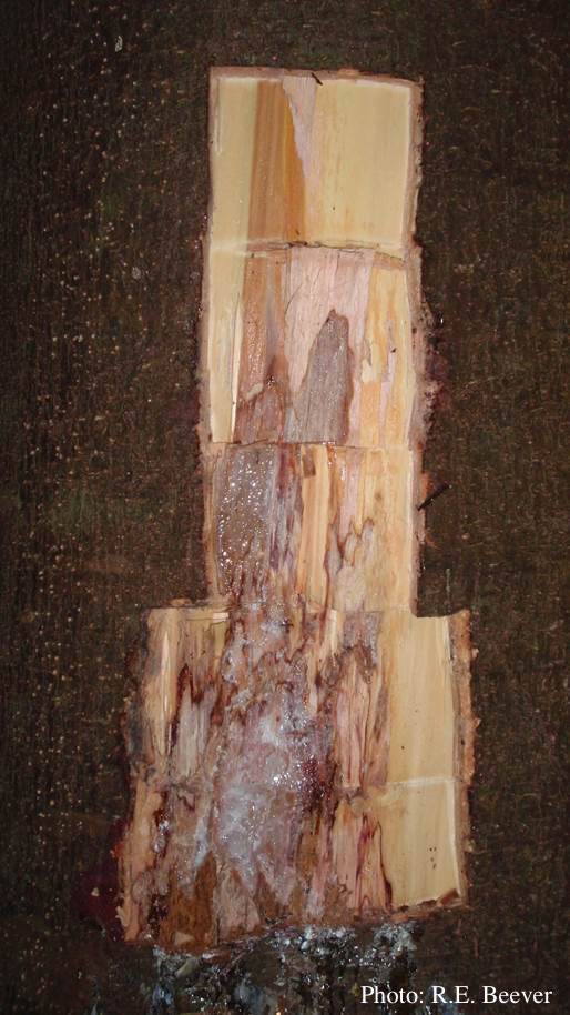 Excavated lesion, with outer bark removed showing extent of disease-front |
|
P. kernoviae leaf wilt 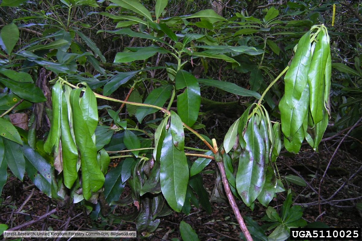 Wilted leaf of infected rhododendron |
P. tentaculata oospores and antheridia  Paragynous antheridium attached to oogonium with oospore |
P. siskiyouensis oogonium with paragynous antheridium  P. siskiyouensis oogonium with paragynous antheridium |