

Crown decline of mature kauri, with branchlets with little or no leaves
Photo Gallery
Site will be retired 9/1/2026
This site is no longer being developed and will be retired on September 1, 2026. Please contact us if you have any questions or would like to provide support to continue the project.
|
P. agathidicia disease symptoms on kauri 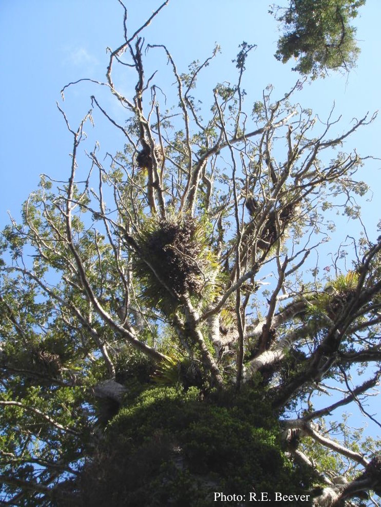 |
P. megasperma colony morphology on PDA  Colony morphology on PDA at 7 days |
P. cambivora oogonium 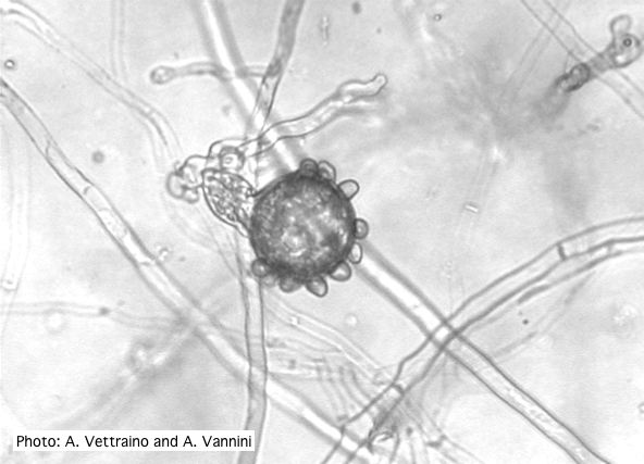 Bullate oogonium with amphyginous antheridium |
|
P. tentaculata microscopic characteristics 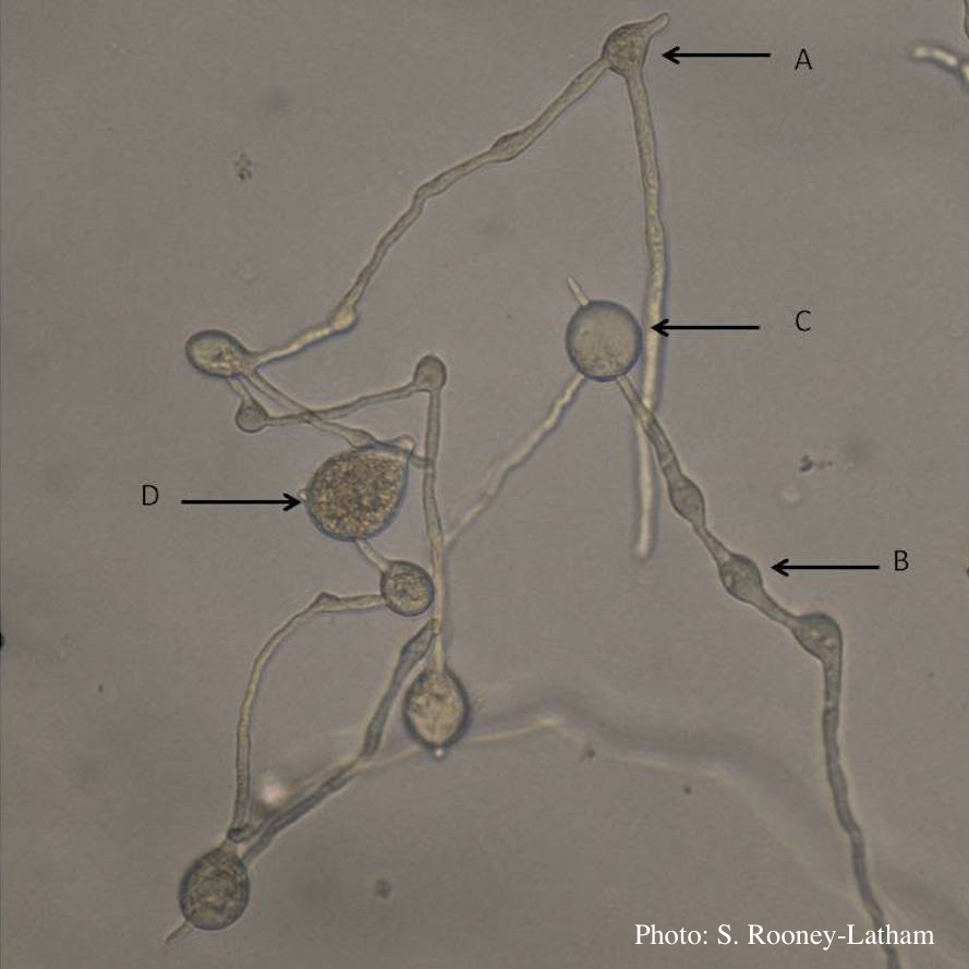 Hyphal swellings occuring at branching points of Mycelium (A), Intercalary hyphal swellings (B), Chlamydospore (C ), Sporangia (D) |
Basal canker on Port-Orford cedar 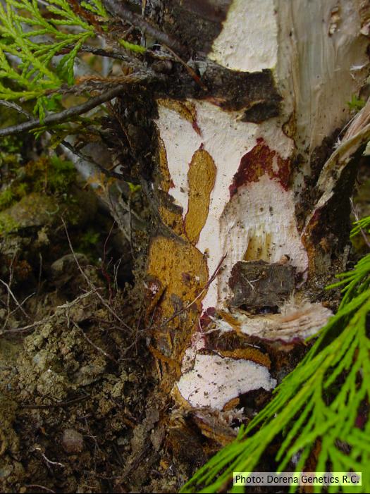 Basal canker on Chamaecyparis lawsoniana |
P. cinnamomi in Australia  Death of Xanthoria and other native vegetation, Victoria, Australia |
|
P. siskiyouensis disease symptoms on Italian alder 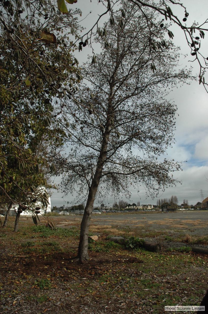 Phytophthora collar rot on Italian alder trees: standing, dead tree |
P. pluvialis on Pinus radiata in New Zealand  Pinus radiata needles showing colour changes following infection with red needle cast disease. The tissues around the initial infection at the base or along the needle senesce, and change yellow and then brown as indicated by the arrows before the needles cast. |
P. pluvialis symptoms on Douglas-fir 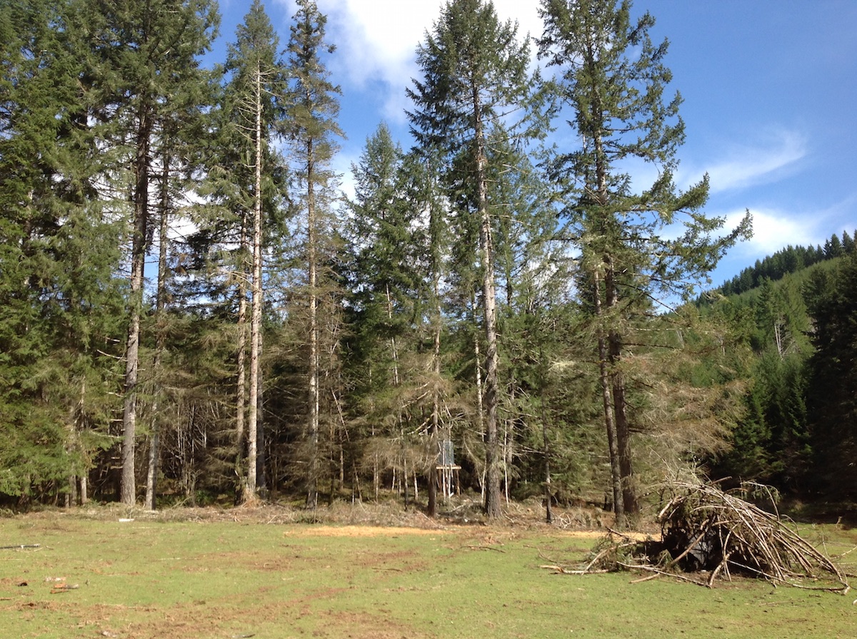 P. pluvialis symptoms of red needle cast on Douglas-fir, western Oregon 2015 |
|
P. ramorum colony morphology on PDA 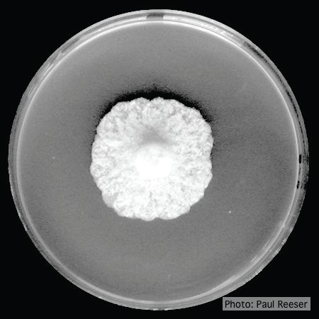 P. ramorum colony morphology on PDA |
P. megakarya sporangia (photo from Q-bank, used with permission). 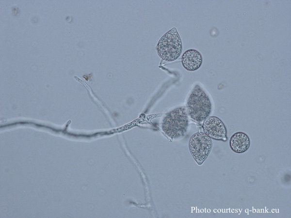 P. megakarya sporangia |
P. katsurae growth morphology on β-CMA 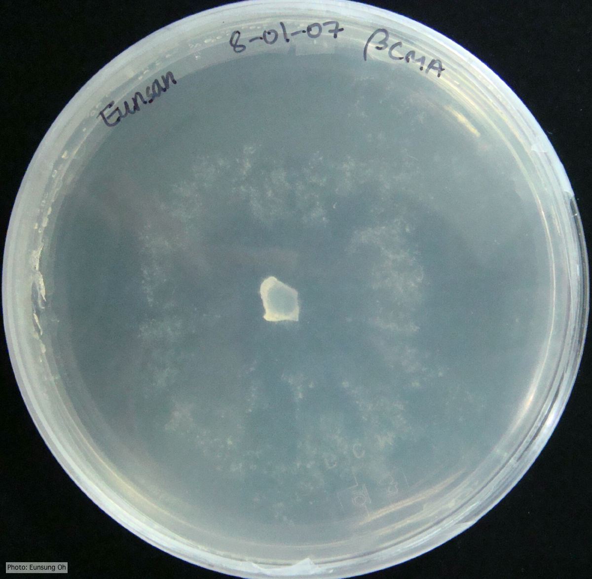 Growth morphology at 7 days on β-CMA |