

P. cambivora oogonium with antheridium
Photo Gallery
Site will be retired 9/1/2026
This site is no longer being developed and will be retired on September 1, 2026. Please contact us if you have any questions or would like to provide support to continue the project.
|
P. cambivora oogonium 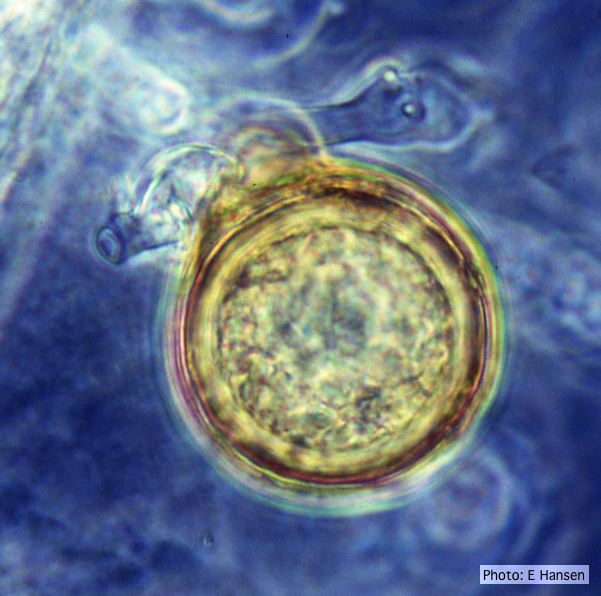 |
P. cambivora disease symptoms 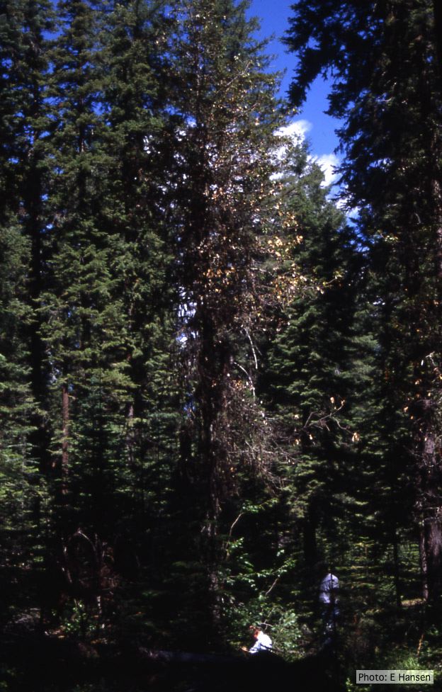 Dead and dying chinquapin infected with P. cambivora |
P. ramorum zoospores 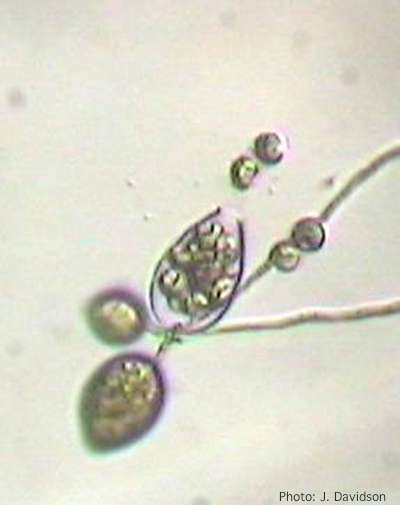 Sporangium of P. ramorum releasing zoospores |
|
P. kernoviae colony morphology  From Mycol.Res 109, 853-859; growth on CA under different conditions |
Comparative gametangial morphology of Phytophthora Clade 5 species  Comparative gametangial morphology of Phytophthora Clade 5 species, with SEM (top) and light microscopy (bottom). P. heveae has smooth walled oogonia with funnel-shaped, amphigynous antheridia. P. agathidicida has mildly stipulate oogonia with globose amphigynous antheridia. P.cocois has mildly bullate oogonia with reflexed amphigynous antheridia. P. castaneae has coarsely bullate oogonium with rugose protuberances and narrow amphigynous antheridia (Weir et al. 2015). |
Vehicle washing  Truck washing to avoid spread of P. lateralis |
|
P. palmivora chlamydospore 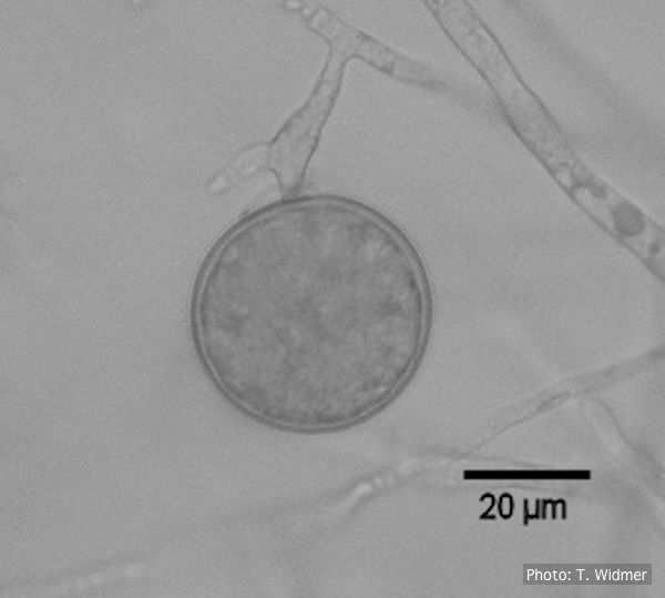 Terminal chlamydospore of P. palmivora |
P. alni symptoms on European Alder  Mature, riparian common alder (A. glutinosa) stand heavily impacted by root and collar rot caused by P. alni |
P. katsurae disease symptoms  Infected chestnut tree with girdling canker on stem |
|
Chlamydospore of P. lateralis 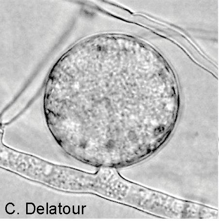 Terminal chlamydospore on a short side stalk |
P. arenaria disease symptoms on Banksia 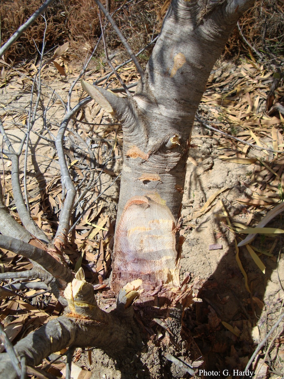 Dead Banksia sp. in a Kwongan heathland on mineral sand near Eneabba, Western Australia recently killed by root and collar rot caused by Phytophthora arenaria |
P. austrocedrae necrotic lesion in phloem 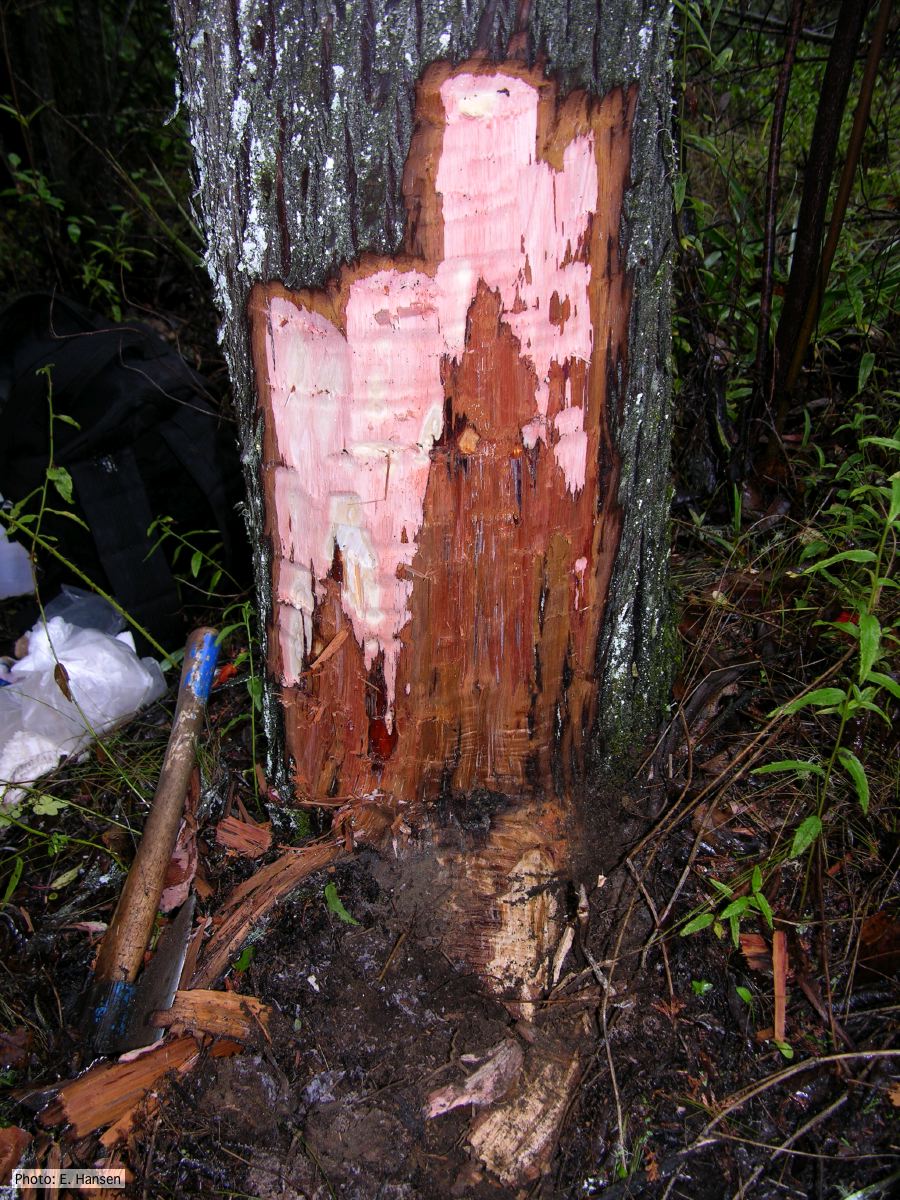 P. austrocedrae - necrotic lesion in phloem |