

Typical decline of Chaemacyparis lawsoniana in Landrévarzec, France
Photo Gallery
Site will be retired 9/1/2026
This site is no longer being developed and will be retired on September 1, 2026. Please contact us if you have any questions or would like to provide support to continue the project.
|
P. lateralis on Port Orford cedar  |
P. megakarya disease symptoms on Theobroma cacao

Symptoms of black pod disease of cocoa (T. cacao)
|
P. pluvialis symptoms on Douglas-fir needles 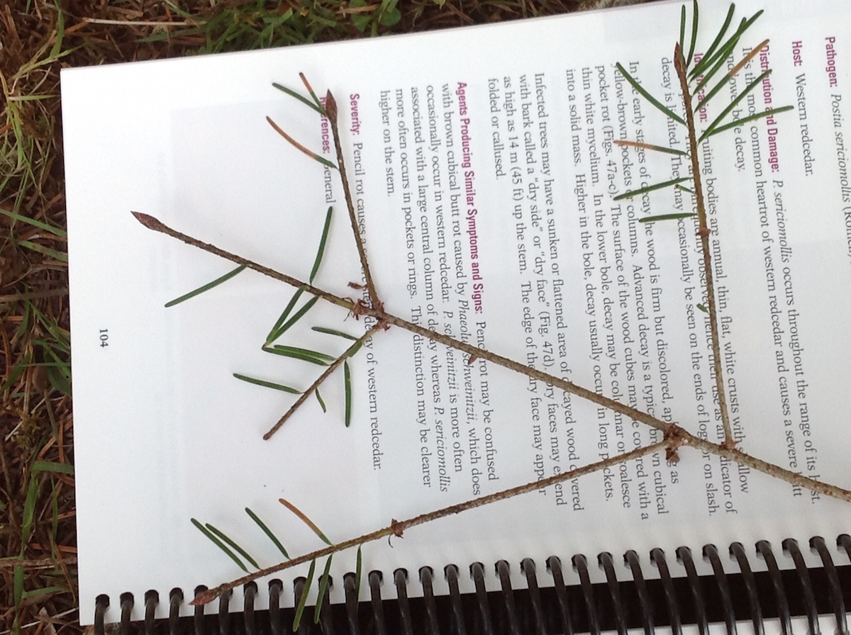 Symptoms of red needle cast on Douglas-fir needles |
|
Phytophthora chlamydospora sporangium 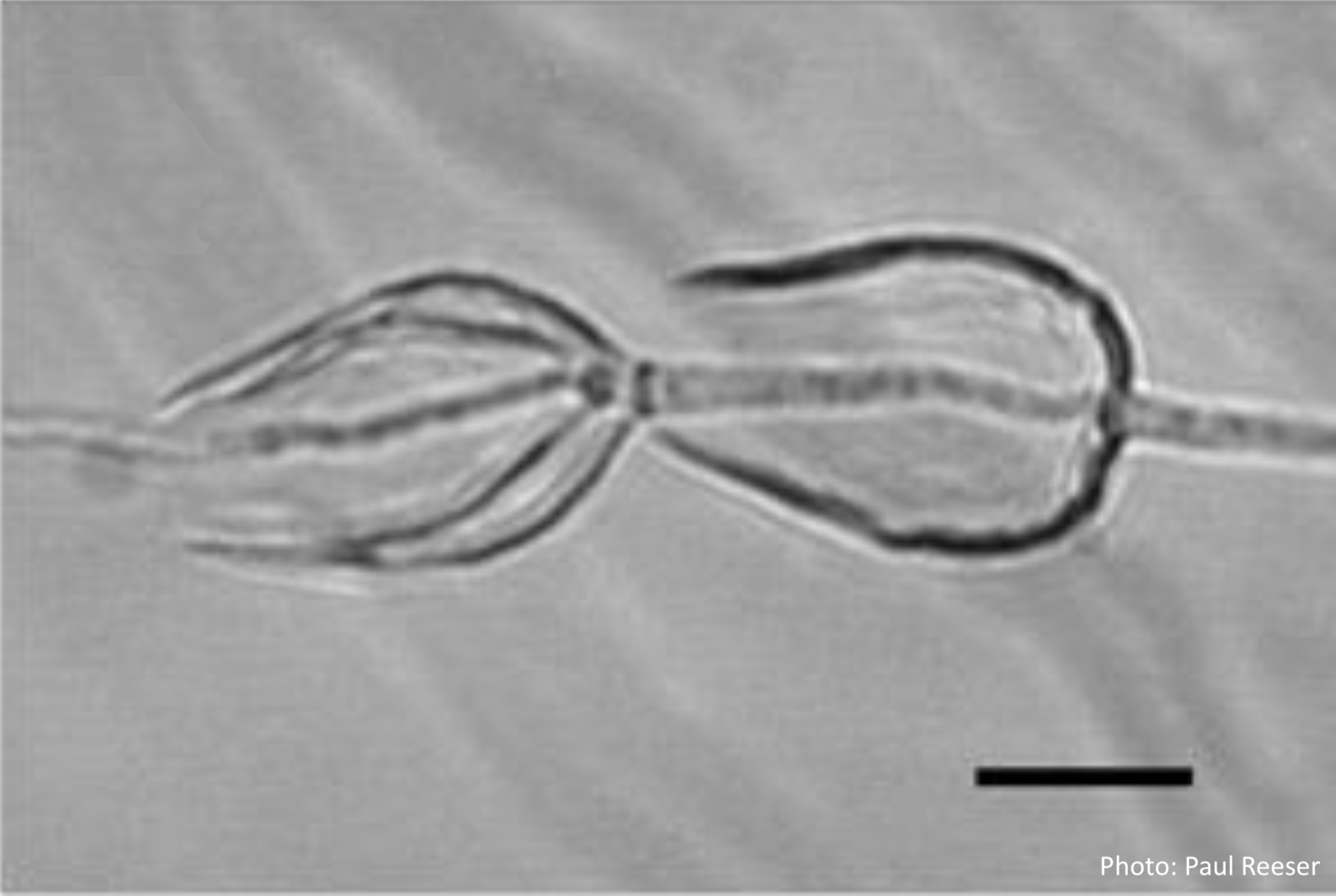 Phytophthora chlamydospora sporangia in water, showing internal proliferation. Bar is 20 µm. |
P. austrocedrae - sporangia drawings 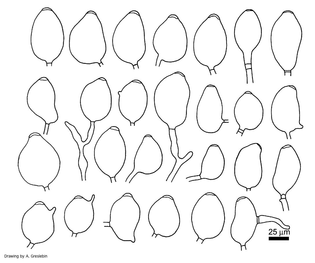 Phytophthora austrocedrae. Morphology of sporangia. Bar: 25 mm. Greslebin et al. 2007 |
P. cambivora disease symptoms 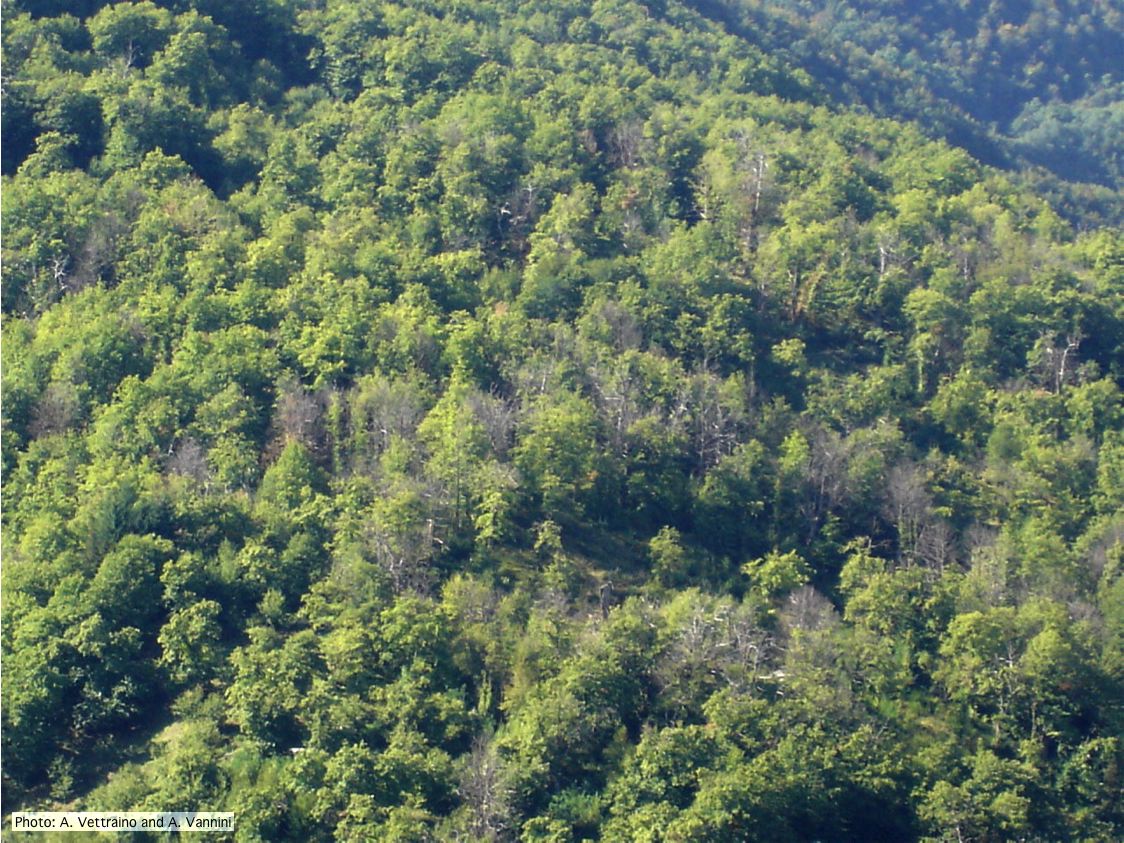 Ink disease impact in sweet chestnut forest in Italy |
|
P. pseudotsugae amphigynous oogonium 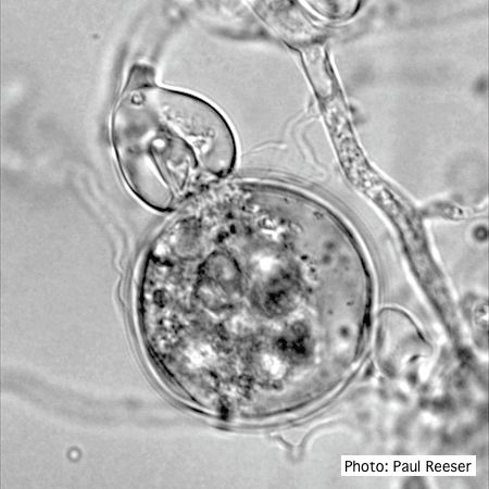 P. pseudotsugae oogonium with amphigynous antheridia |
P. kernoviae oogonia 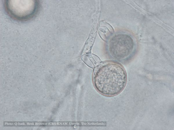 Oogonium with amphigynous antheridia, photo from Q-bank, used with permission |
P. pinifolia hyphal swellings  Spherical hyphal swelling with radiating hyphae (from Duran et al. 2008). Scale bar = 20 μm. |
|
P. arenaria sporangia  Globose papillate sporangia of Phytophthora arenaria on V8 agar flooded with soil extract. (Scale bar = 20 μm) |
P. pluvialis symptoms on Douglas-fir 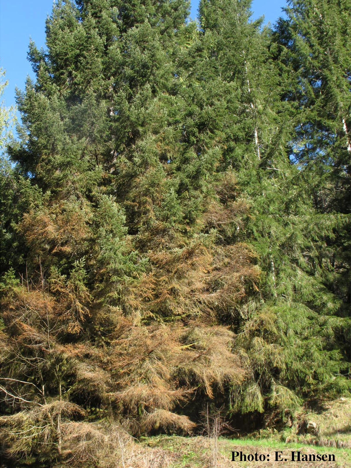 Red needle cast symptoms on Douglas-fir in western Oregon, 2015 |
P. nemorosa oogonium 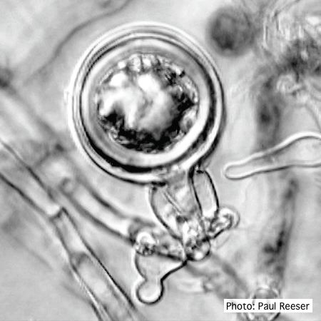 Oogonium with amphigynous antheridium |