

P. cambivora oogonium with antheridium
Photo Gallery
|
P. cambivora oogonium 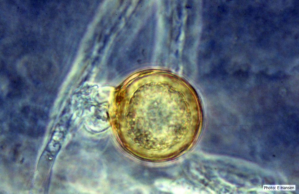 |
P. cambivora colony morphology on PDA 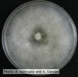 Uniform fluffy colony morphology at 14 days at 20°C on PDA |
P. pinifolia sporulation 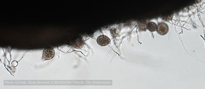 Sporulation on edge of hemp seed, photo from Q-bank, used with permission. |
|
P. cinnamomi cork oak decline  Cork oak decline, Portugal |
P. pinifolia sporangium 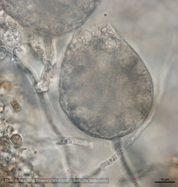 Cysts remain in sporangium after discharge, photo from Q-bank, used with permission |
P. frigida oogonium  Oogonium and oospore with amphigynous antheridium |
|
P. arenaria disease symptoms on Banksia 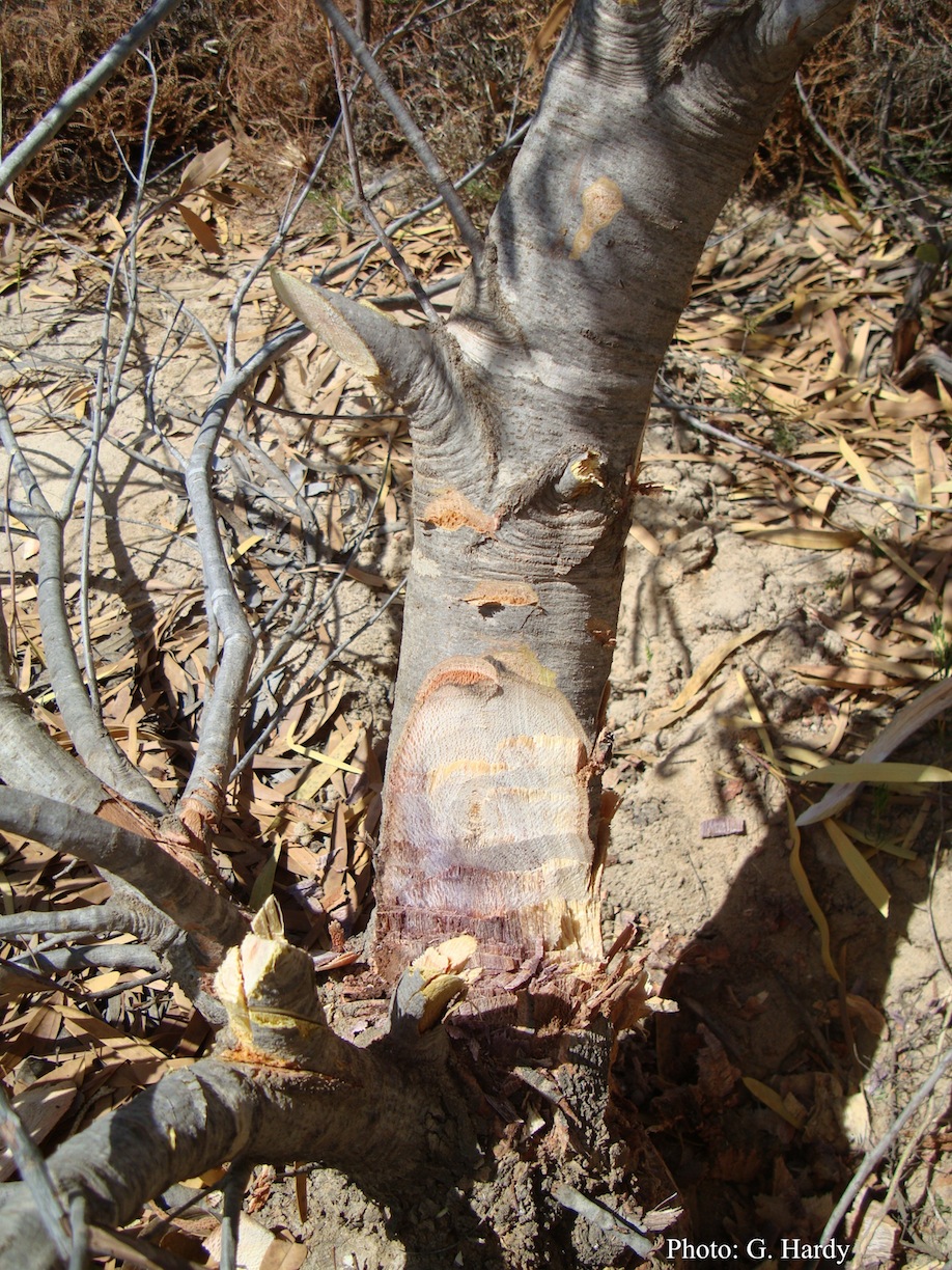 Dead Banksia sp. in a Kwongan heathland on mineral sand near Eneabba, Western Australia recently killed by root and collar rot caused by Phytophthora arenaria |
P. tentaculata disease symptoms on California mugwort 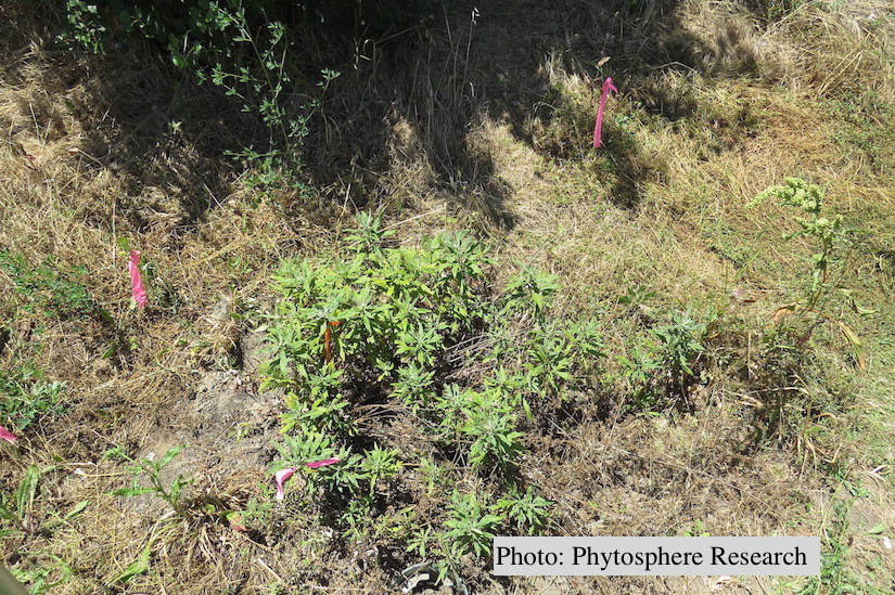 Outplanted California mugwort (Artemisia douglasiana) infected with P. tentaculata, 4.5 years after planting. Plant shows stunting and chlorosis. (P. cryptogea and P. lacustris were also baited from roots/soil of this plant). |
P. katsurae oogonium  Micrograph of warty oogonium |
|
P. alni symptoms on European Alder 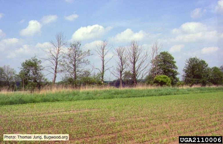 Mature, riparian common alder (A. glutinosa) stand with high impact of Phytophthora root and collar rot. |
Growth of P. arenaria on MEA 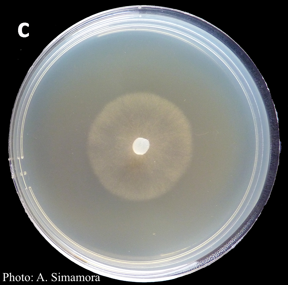 Colony morphology of Phytophthora arenaria after 7 days at 20°C on malt extract agar |
P. ramorum chlamydospores 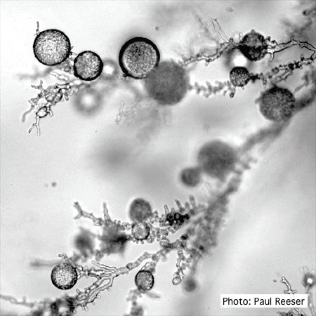 P. ramorum chlamydospores |