

P. chlamydospora colony morphology on V8 agar
Photo Gallery
Site will be retired 9/1/2026
This site is no longer being developed and will be retired on September 1, 2026. Please contact us if you have any questions or would like to provide support to continue the project.
|
P. chlamydospora colony morphology on V8 agar 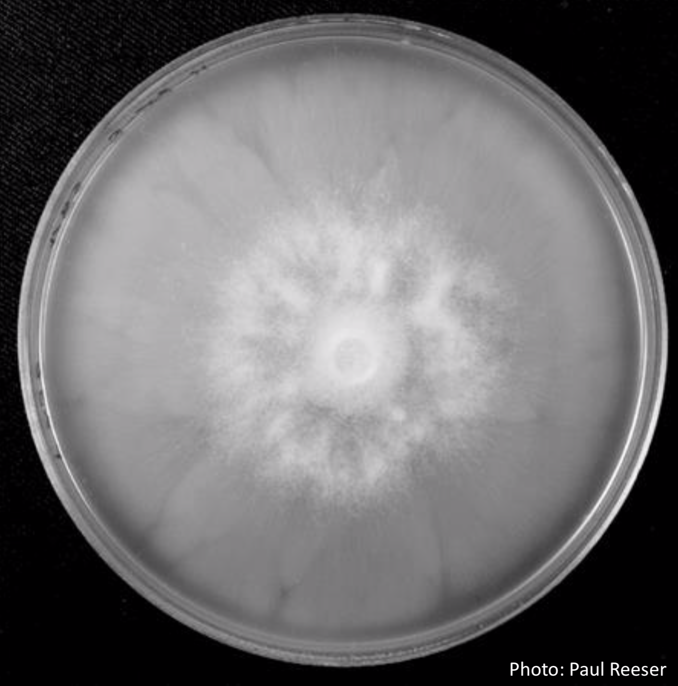 |
P. katsurae disease symptoms 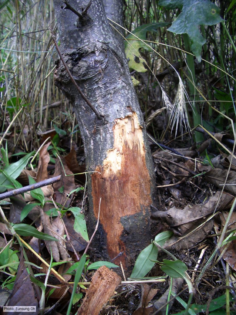 Infected chestnut tree with girdling canker on stem |
P. katsurae disease symptoms 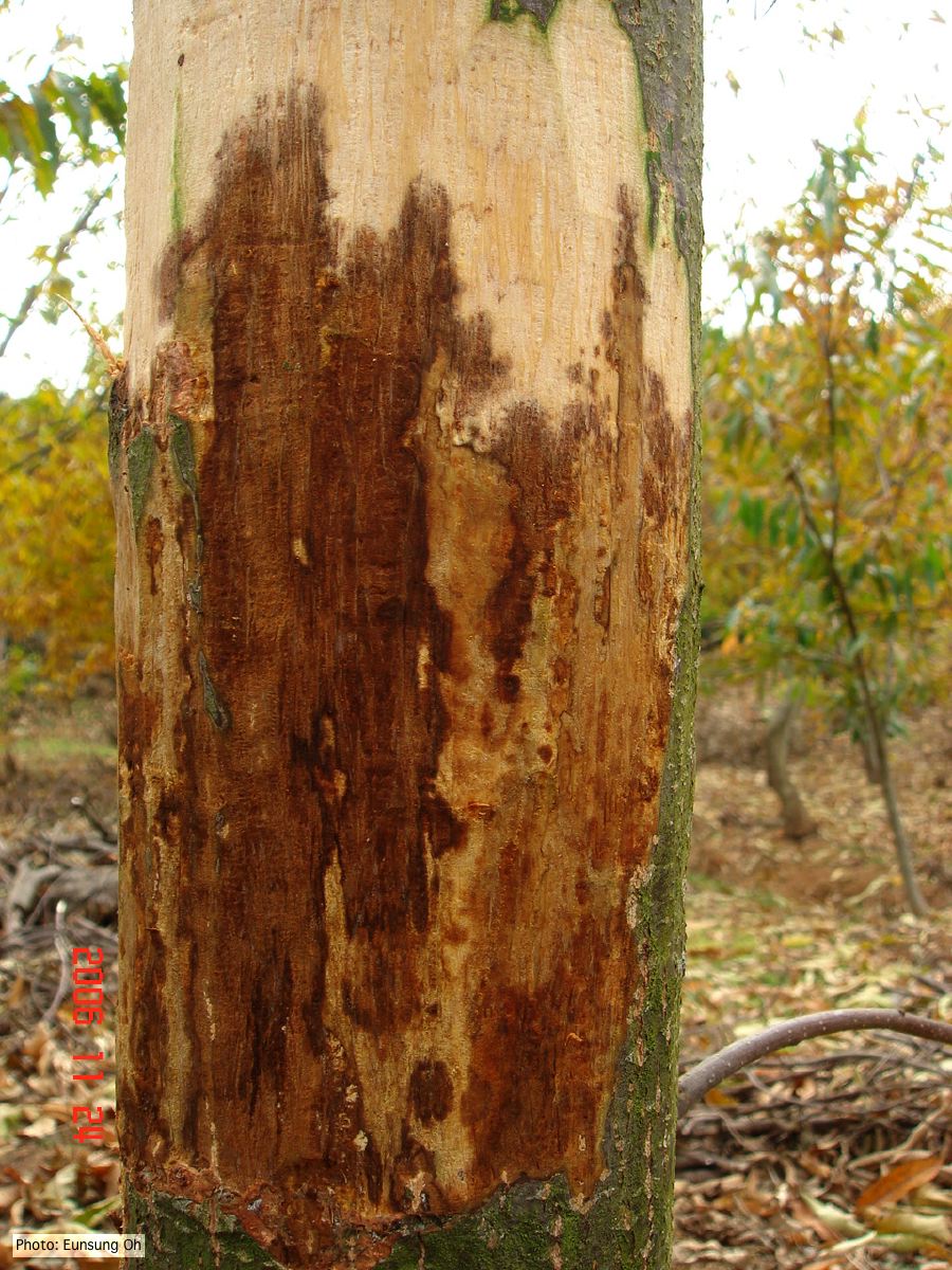 Infected chestnut (Castanea) with girdling canker |
|
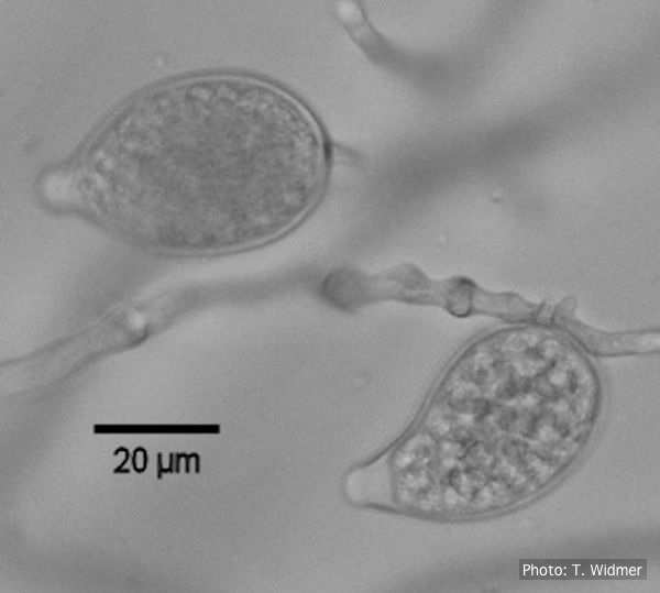
P. cactorum sporangia 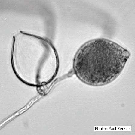 Broadly ovoid, papillate sporangia in water. |
P. ramorum canker 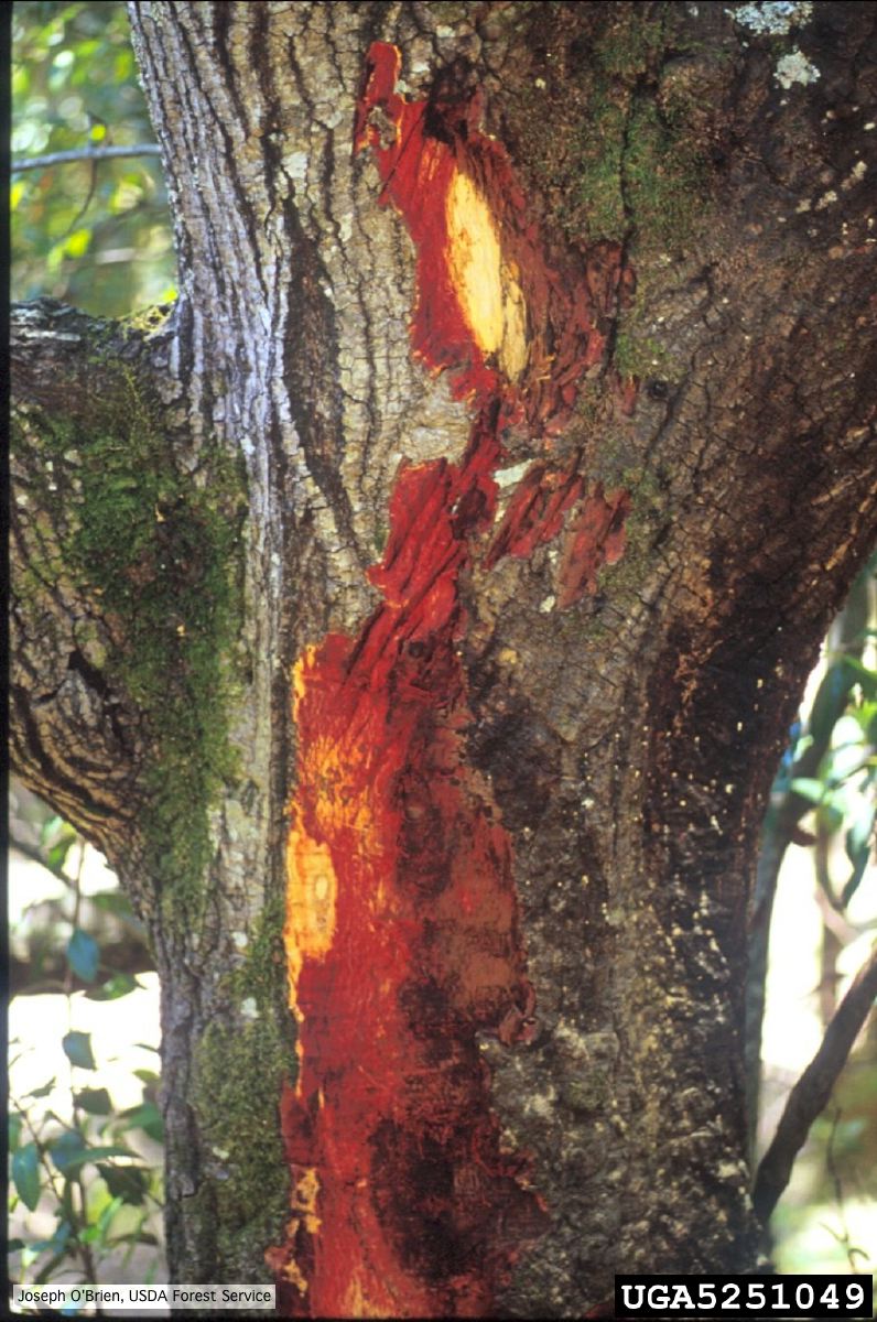 Bark discoloration and zone lines in coast live oak (Quercus agrifolia) |
P. palmivora sporangia
 P. palmivora caducous papillate sporangia
|
|
Phytophthora taxon Agathis bole canker  Canker on a Kauri tree, New Zealand |
P. pinifolia coenocytic hyphae 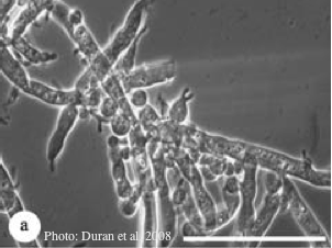 Coenocytic hyphae (from Duran et al. 2008). Scale bar = 20 μm. |
P. pseudotsugae sporangium 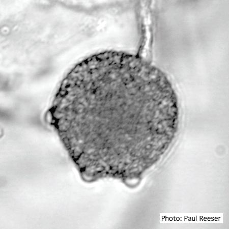 Broadly ovoid, papillate sporangium in water |
|
P. cactorum oogonium 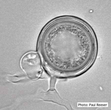 Oogonium with paragynous antheridia close to oogonial stalk. Oospores are slightly aplerotic. |
P. austrocedrae semipapillate sporangium  P. austrocedrae - semipapillate sporangium with off-center attachment. |
P. cactorum colony morphology on PDA 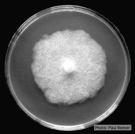 Colony morphology on PDA at 14 days |