

Disease symptoms on a cocoa pod
Photo Gallery
Site will be retired 9/1/2026
This site is no longer being developed and will be retired on September 1, 2026. Please contact us if you have any questions or would like to provide support to continue the project.
|
P. megakarya disease symptoms on Theobroma cacao fruit 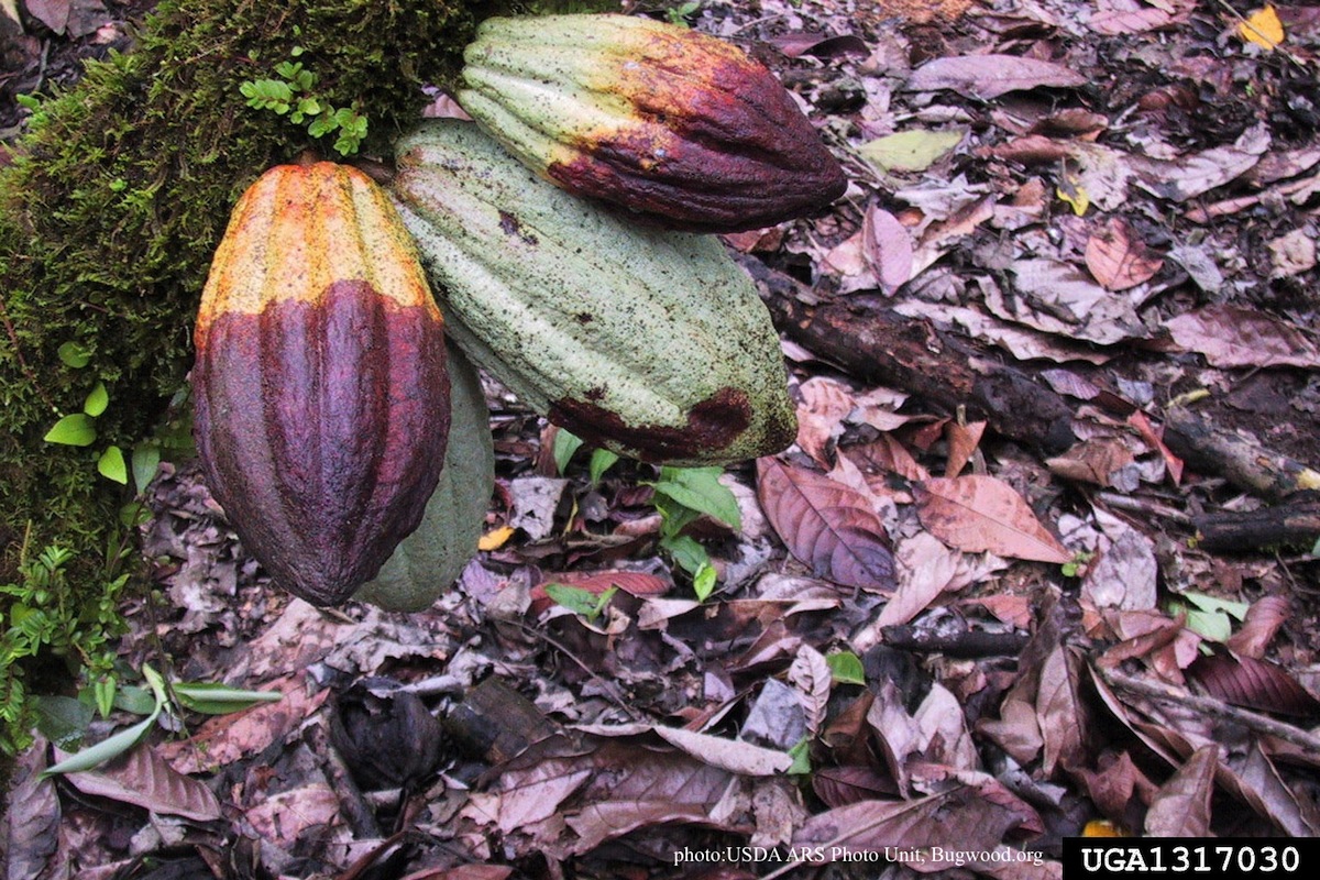 |
Growth of P. megakarya on PDA 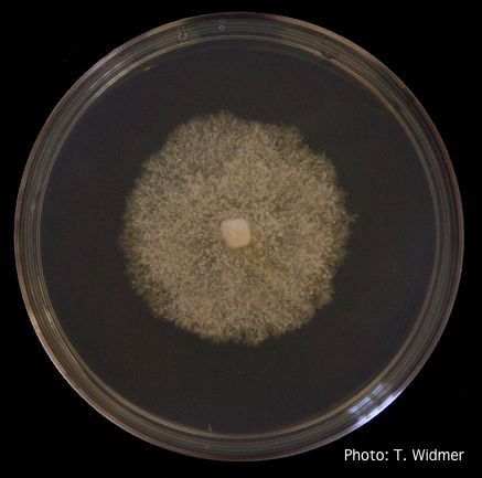 Growth of P. megakarya on potato dextrose agar |
P. tentaculata disease symptoms on California mugwort 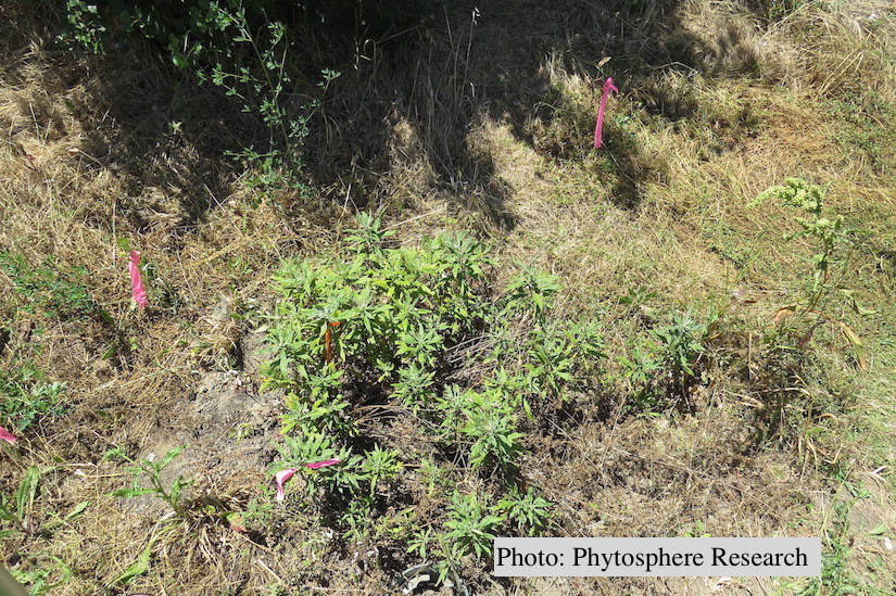 Outplanted California mugwort (Artemisia douglasiana) infected with P. tentaculata, 4.5 years after planting. Plant shows stunting and chlorosis. (P. cryptogea and P. lacustris were also baited from roots/soil of this plant). |
|
P. palmivora oogonium 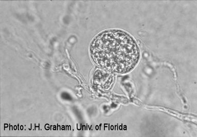 P. palmivora oogonium |
P. ramorum canker 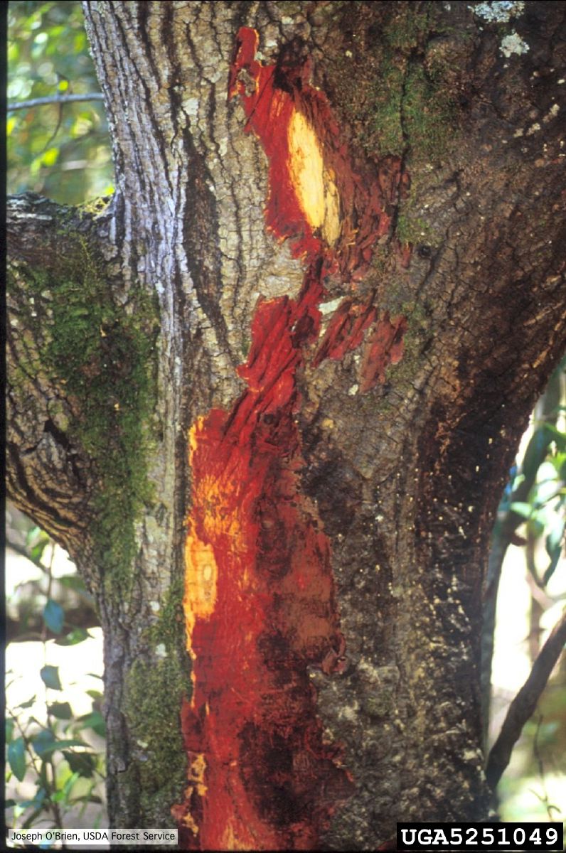 Bark discoloration and zone lines in coast live oak (Quercus agrifolia) |
P. cambivora sporangium 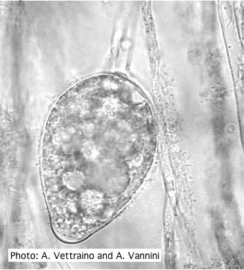 Ovoid non-papillate sporangia with well-rounded base |
|
P. pinifolia colony morphology on CMA-NARP 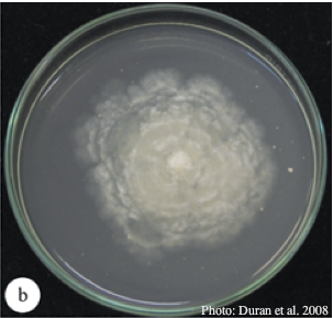 Colony morphology of P. pinifolia at 20°C on CMA-NARP after 3 weeks. From Duran et al. 2008 |
P. arenaria oogonium  Aplerotic oogonia of P. arenaria with paragynous antheridia. Scale bar = 20 μm |
P. alni oogonium 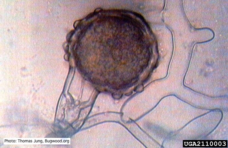 Bullate oogonium of P. alni (German variant) with oospore and amphigynous antheridium. |
|
P. ramorum sporangium 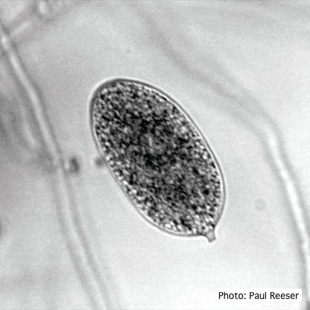 P. ramorum sporangium |
P. alni basal canker on European Alder 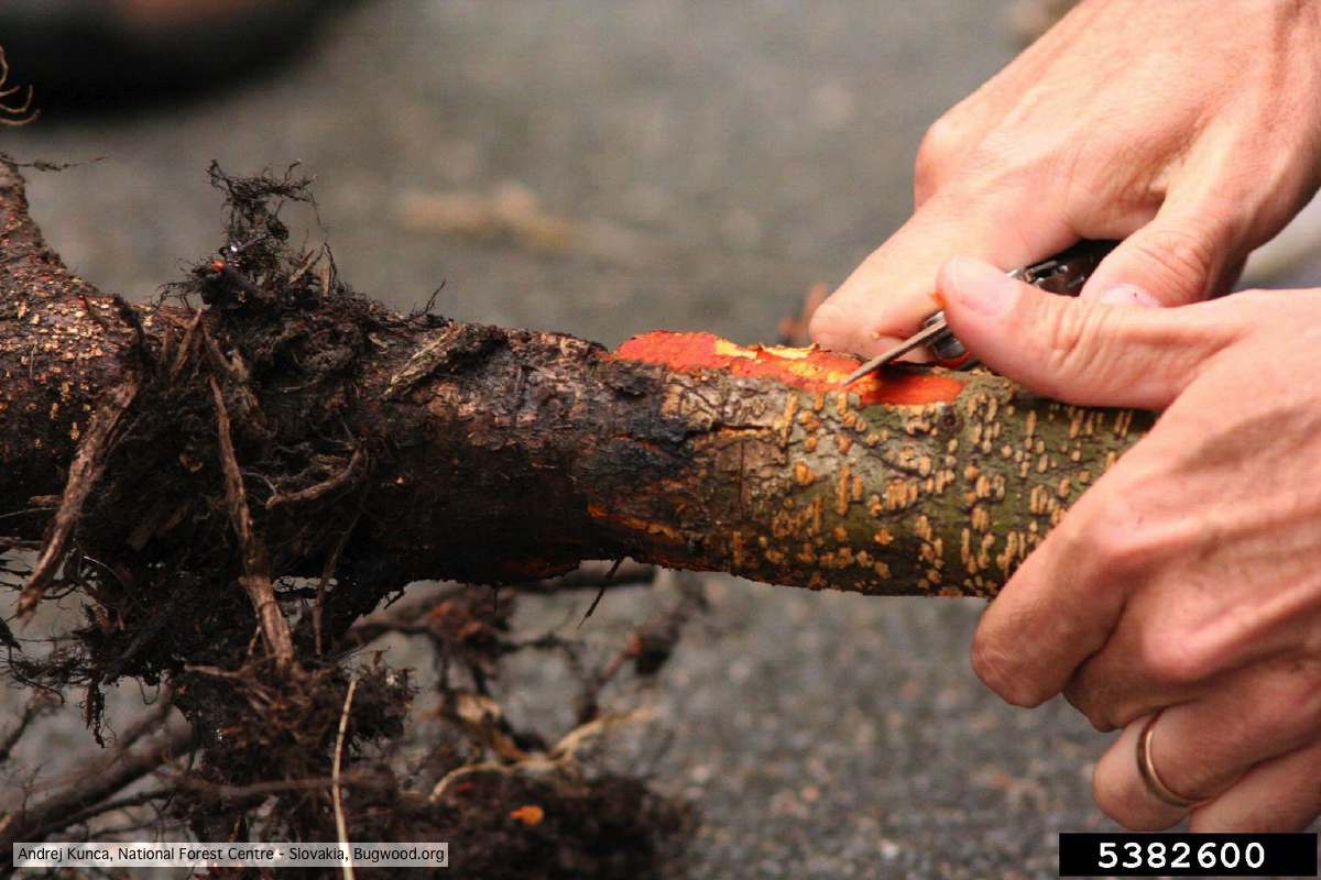 P. alni basal canker on European Alder (Alnus glutinosa) |
P. ramorum sporangia and chlamydospores 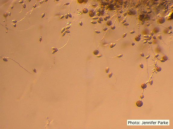 Sporangia and chlamydospores of P. ramorum |