

Colony morphology of Phytophthora arenaria after 7 days at 20°C on carrot agar
Photo Gallery
|
Growth of P. arenaria on CA  |
P. ramorum Dieback and shoot blight on Grand fir 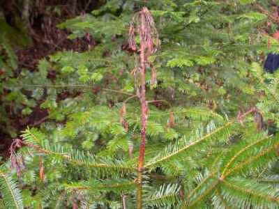 Dieback and shoot blight symptoms caused by P. ramorum on Abies grandis |
P. ramorum colony morphology on CMA PARP 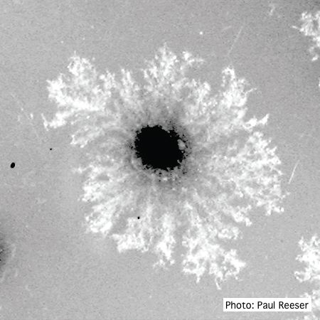 P. ramorum colony morphology on CMA PARP |
|
P. cactorum sporangia  P. cactorum sporangia |
P. siskiyouensis disease symptoms on Italian alder 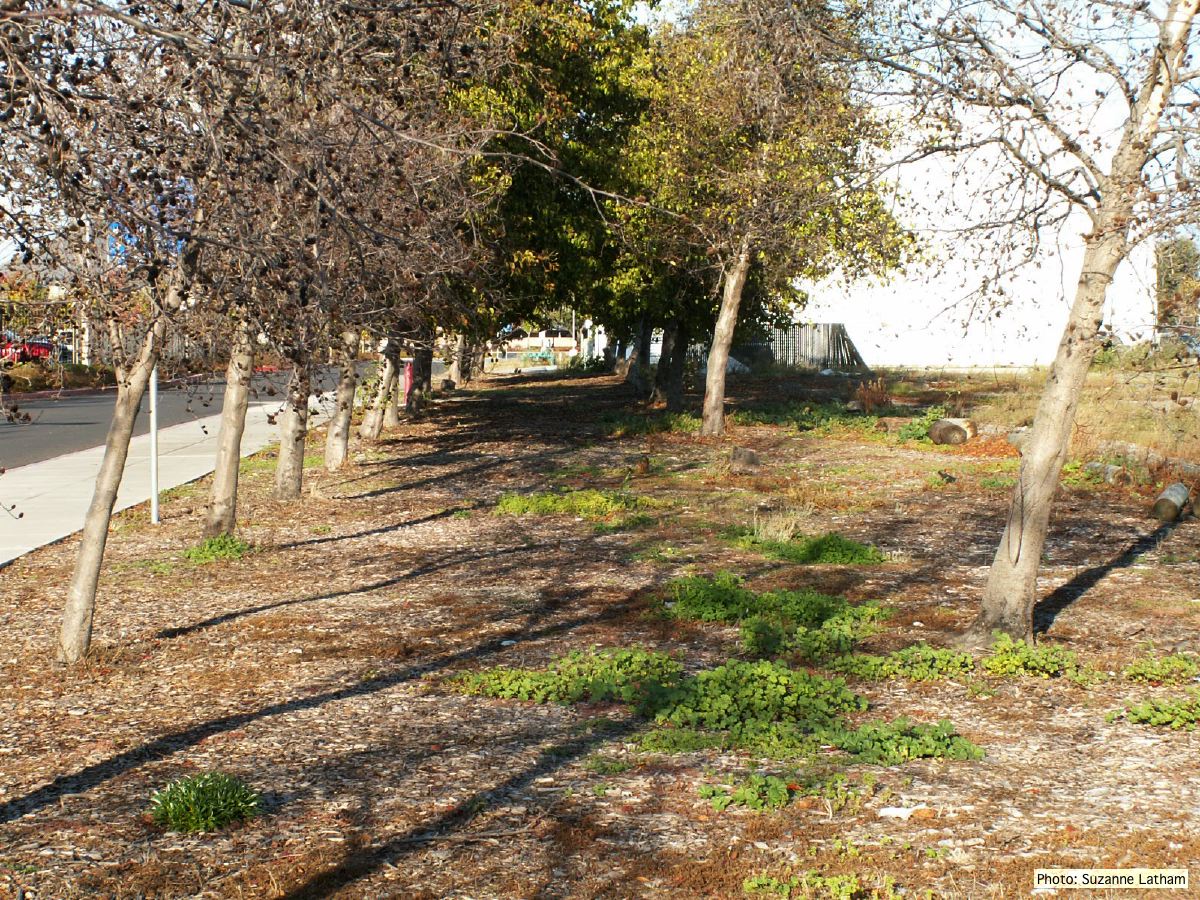 Grove of dying trees in a commercial landscape in Foster City, CA |
P. cinnamomi cork oak decline  Red oak with embedded lesions, France |
|
P. kernoviae canker  Bole lesion on Fagus sylvatica |
P. frigida symptoms 3 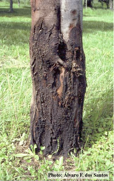 Black wattle bark with symptoms of gummosis |
P. tentaculata disease symptoms on California mugwort 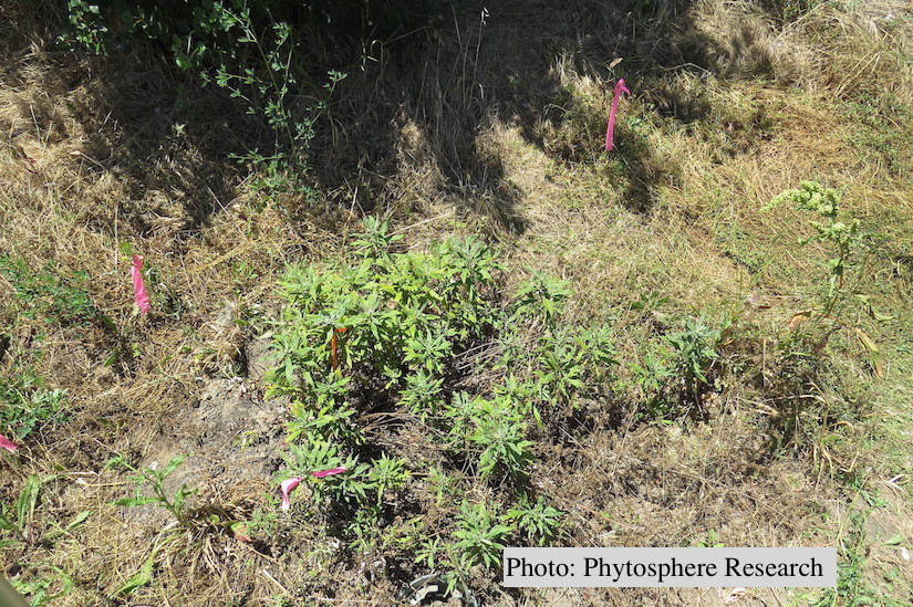 Outplanted California mugwort (Artemisia douglasiana) infected with P. tentaculata, 4.5 years after planting. Plant shows stunting and chlorosis. (P. cryptogea and P. lacustris were also baited from roots/soil of this plant). |
|
P. pinifolia colony morphology on V8 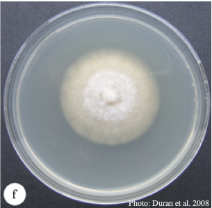 Colony morphology of P. pinifolia at 20°C on V8 after 3 weeks. From Duran et al. 2008 |
P. pinifolia coenocytic hyphae 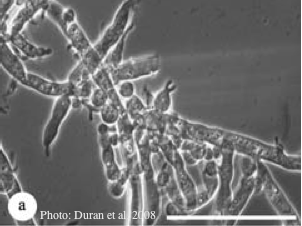 Coenocytic hyphae (from Duran et al. 2008). Scale bar = 20 μm. |
P. frigida oogonium  Oogonium and oospore with amphigynous antheridium |