

Ovoid, semi-papillate sporangia showing sympodial development of sporangiophore
Photo Gallery
Site will be retired 9/1/2026
This site is no longer being developed and will be retired on September 1, 2026. Please contact us if you have any questions or would like to provide support to continue the project.
|
P. nemorosa sporangia  |
P. pluvialis hyphal swellings 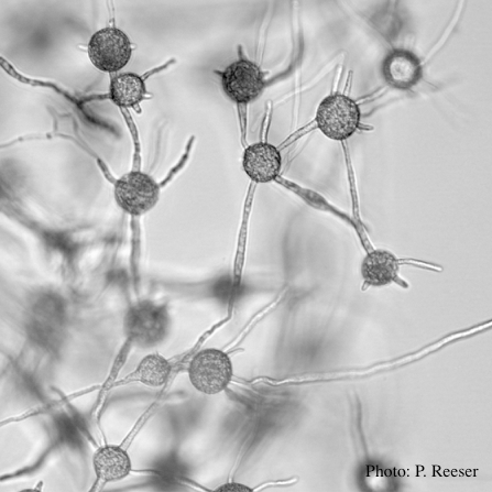 P. pluvialis hyphal swellings in water |
P. pinifolia on Pinus radiata 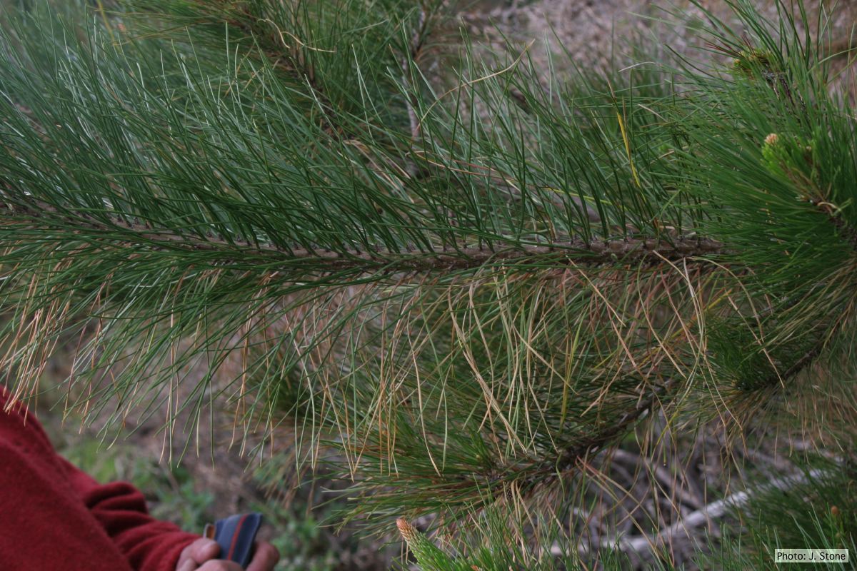 Dead needles on lower side of P. radiata branch. |
|
P. pseudosyringae sporangium 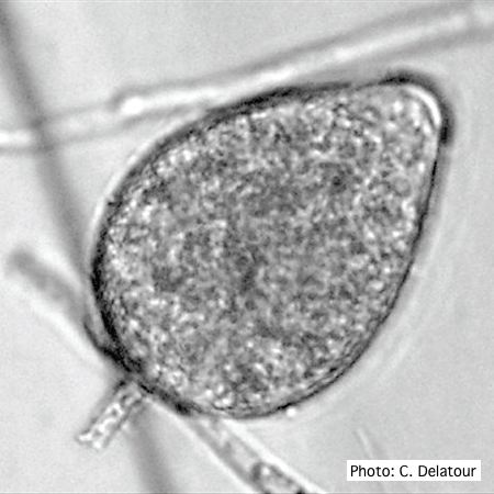 Ovoid, semipapillate sporangia showing medium length pedicel |
P. cambivora oogonium  Bullate oogonium and and two-celled amphigynous antheridium |
P. cryptogea colony morpholgy on PDA 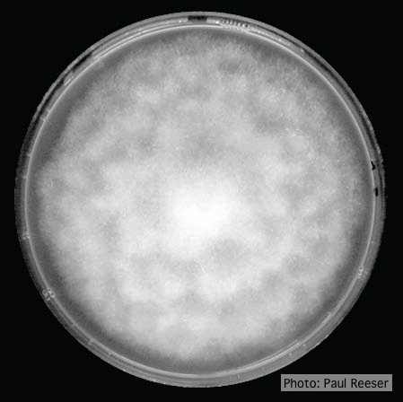 Colony morphology on PDA at 14 days |
|
Growth of P. palmivora on CMA 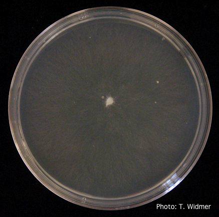 Growth of P. palmivora on corn meal agar |
P. boehmeriae chlamydospore 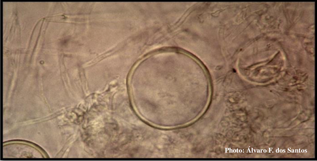 Globose chlamydospore of P. boehmeriae |
P. tentaculata disease symptoms on California mugwort  Nursery grown California mugwort plant (Artemisia douglasiana) infected with P. tentaculata and exhibiting severe root and crown rot |
|
P. siskiyouensis sporangia 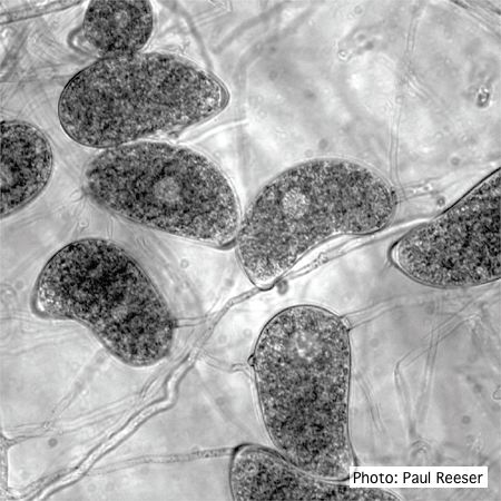 Sporangia showing a variety of shapes and orientations of semi-papillae and sporangiophores |
P. cinnamomi hyphal swellings 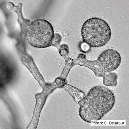 P. cinnamomi hyphal swellings (or thin walled chlamydospores) |
P. cambivora tar spots 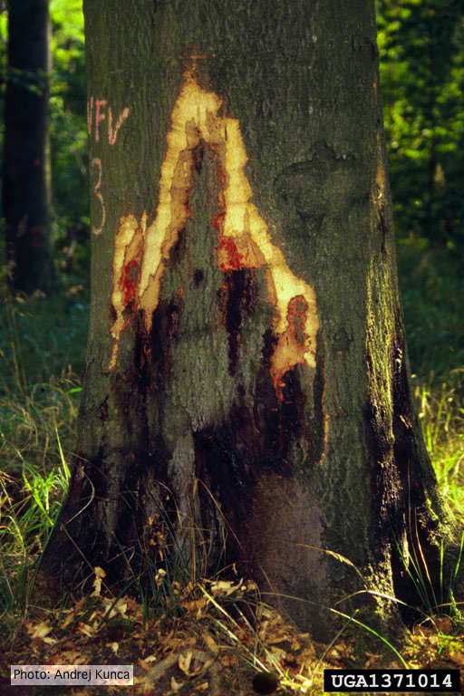 Tar spots on European beech (Fagus sylvatica) with bark removed. Lesse, Germany |