

Phytophthora collar rot on Italian alder trees: standing, dead tree
Photo Gallery
Site will be retired 9/1/2026
This site is no longer being developed and will be retired on September 1, 2026. Please contact us if you have any questions or would like to provide support to continue the project.
|
P. siskiyouensis disease symptoms on Italian alder 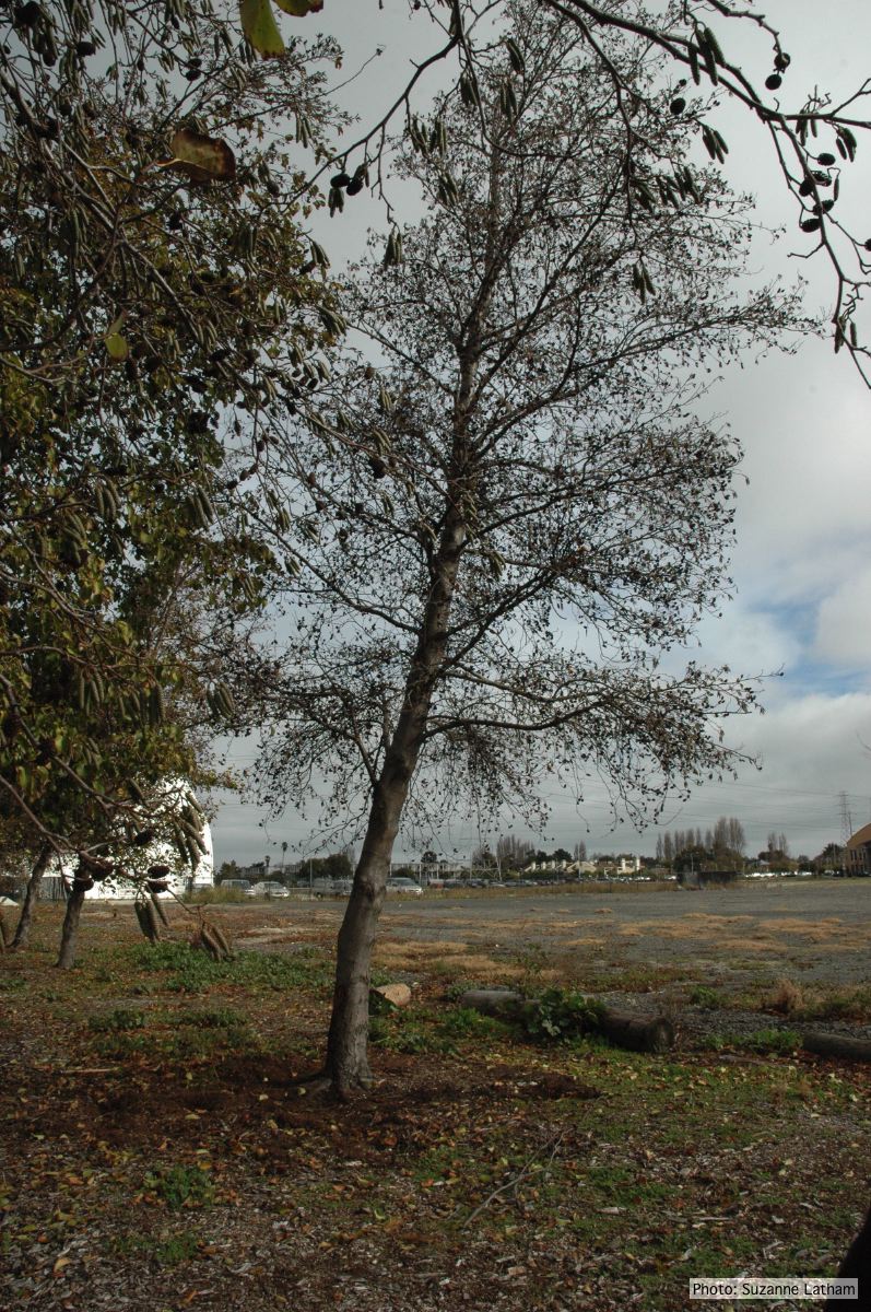 |
Basal canker on Port-Orford cedar 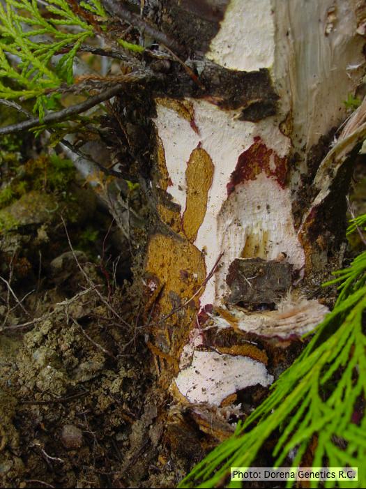 Basal canker on Chamaecyparis lawsoniana |
P. pluvialis symptoms on Douglas-fir 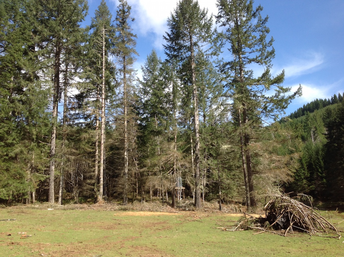 P. pluvialis symptoms of red needle cast on Douglas-fir, western Oregon 2015 |
|
P. kernoviae colony morphology on CMA PARPH 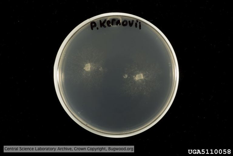 Organism grown on CMA PARP[H]; Plant disease 70, 1038-1043 |
Dying Port Orford Cedar trees  |
Dead Port Orford Cedar trees 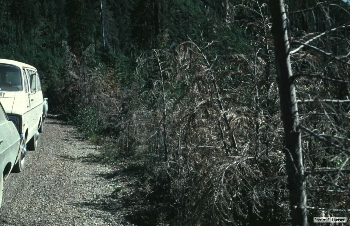 Dead Chamaecyparis lawsoniana trees along road |
|
P. cinnamomi hyphal swelling 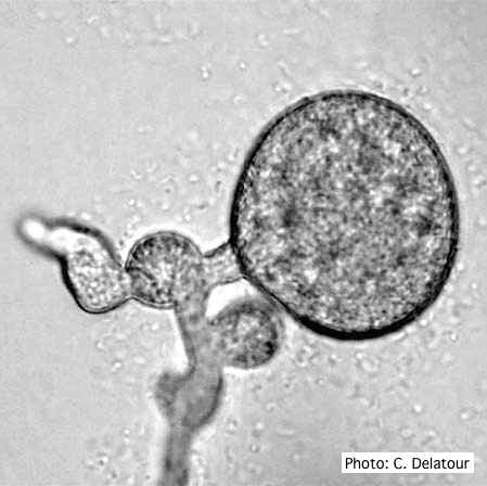 P. cinnamomi hyphal swelling (or thin walled chlamydospores) |
P. pinifolia colony morphology on CMA-NARP 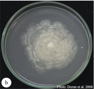 Colony morphology of P. pinifolia at 20°C on CMA-NARP after 3 weeks. From Duran et al. 2008 |
Black pod disease of cacao caused by P. palmivora 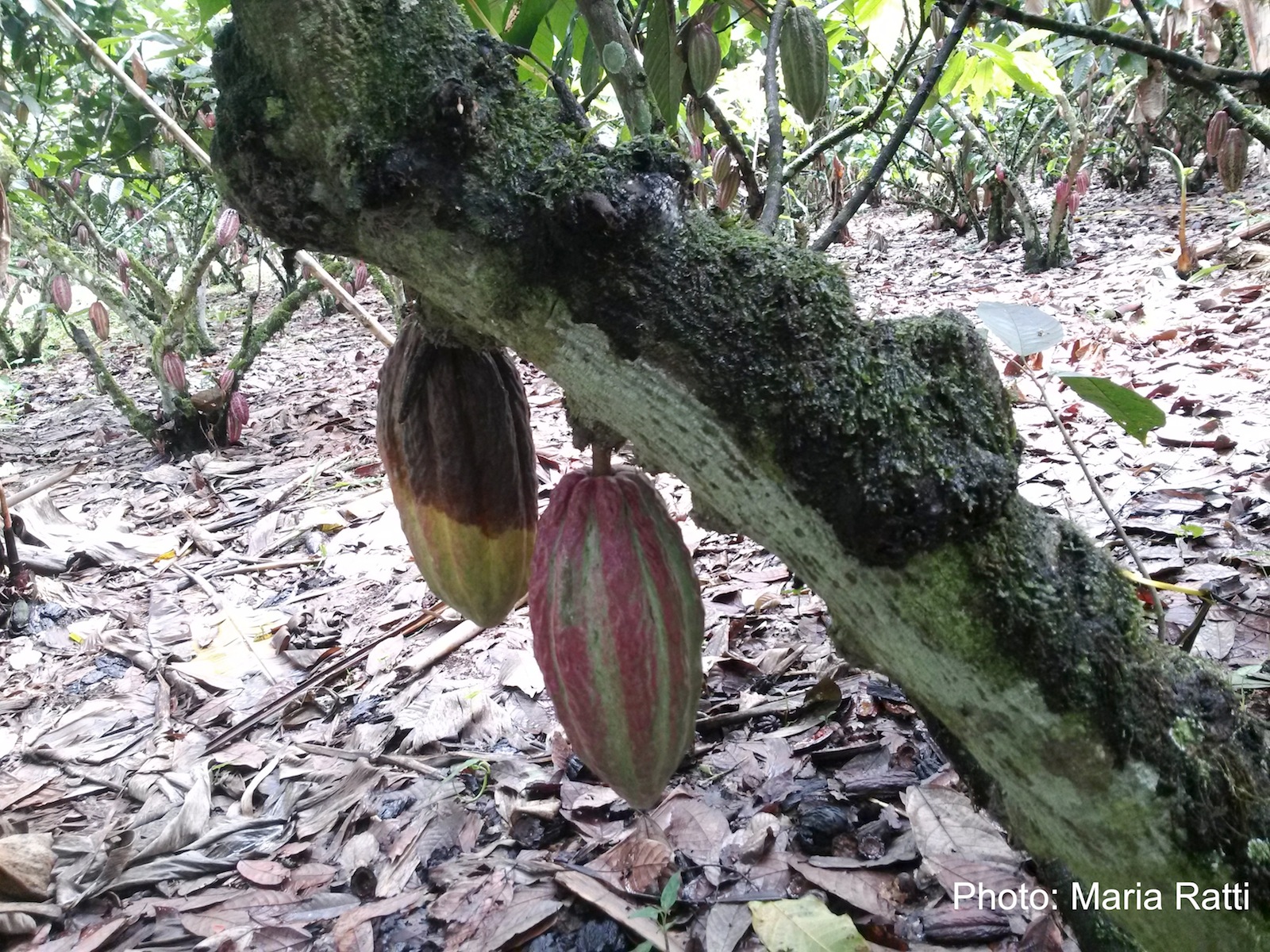 Black pod of cacao in Ecuador caused by P. palmivora (see lesioned fruit on left). |
|
P. boehmeriae chlamydospore 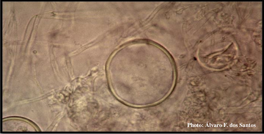 Globose chlamydospore of P. boehmeriae |
P. cryptogea colony morpholgy on V8  Colony morphology on V8 at 14 days |
P. arenaria oogonium  Aplerotic oogonia of P. arenaria with paragynous antheridia. Scale bar = 20 μm |