Terminal chlamydospore of P. megakarya


|
P. megakarya chlamydospore 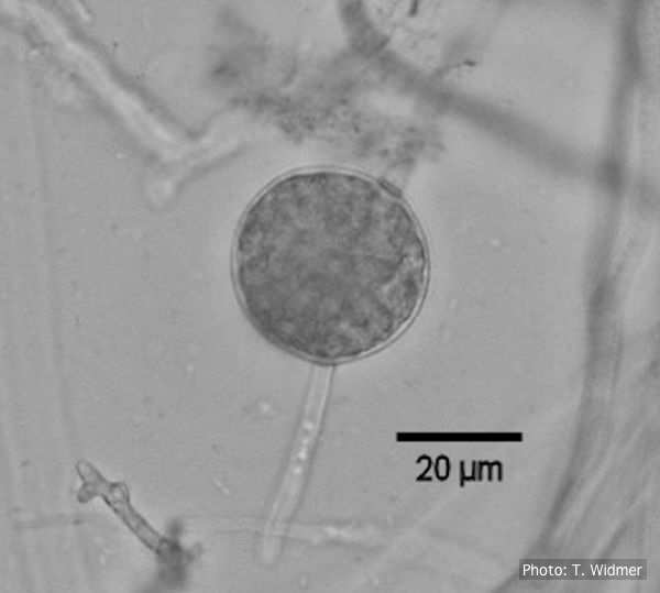
Terminal chlamydospore of P. megakarya
|
P. chlamydospora colony morphology on V8 agar 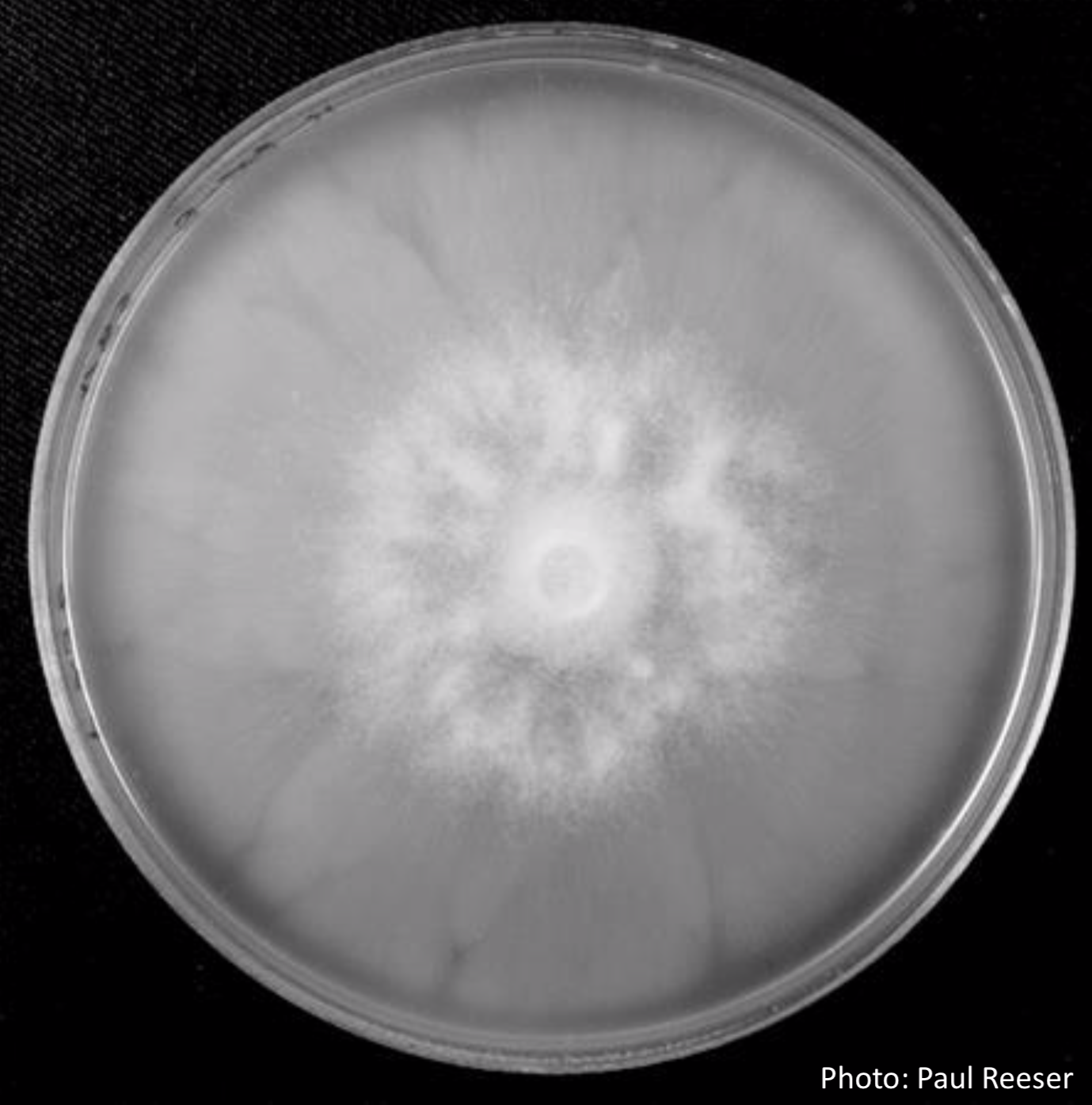 P. chlamydospora colony morphology on V8 agar |
P. alni lesion in alder, Illwald, France 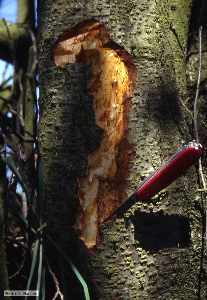 P. alni lesion in alder, Illwald, France |
|
P. agathidicia sporangia 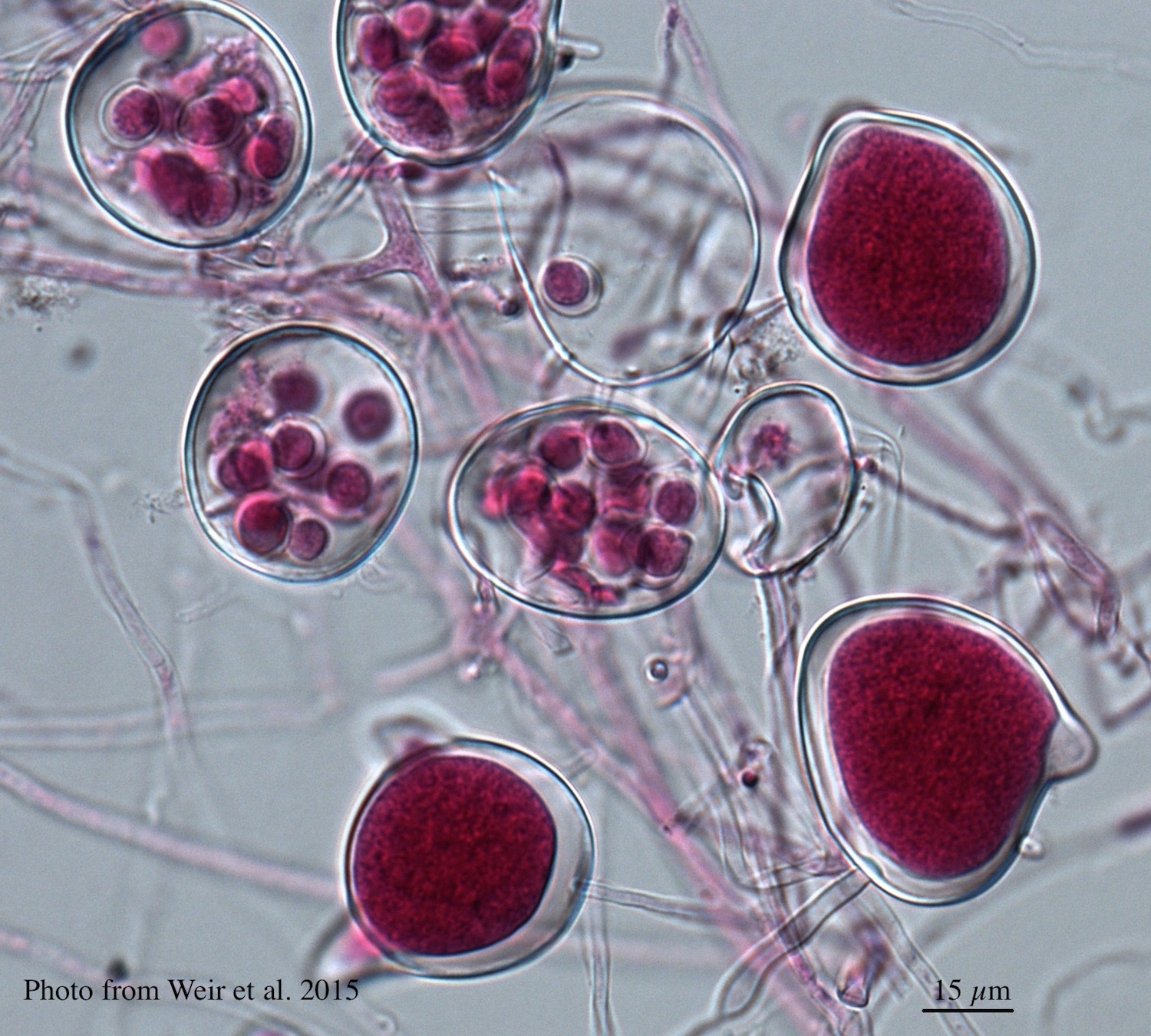 Differentiation of the cytoplasm within papillate sporangia into acid fuchsin stained zoospores |
P. siskiyouensis disease symptoms on Italian alder 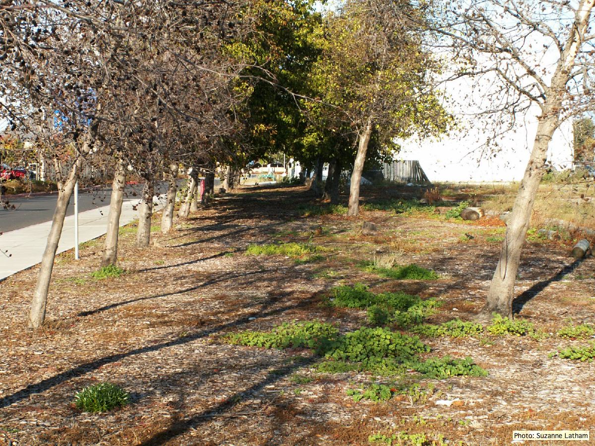 Grove of dying trees in a commercial landscape in Foster City, CA |
P. arenaria disease symptoms on Banksia 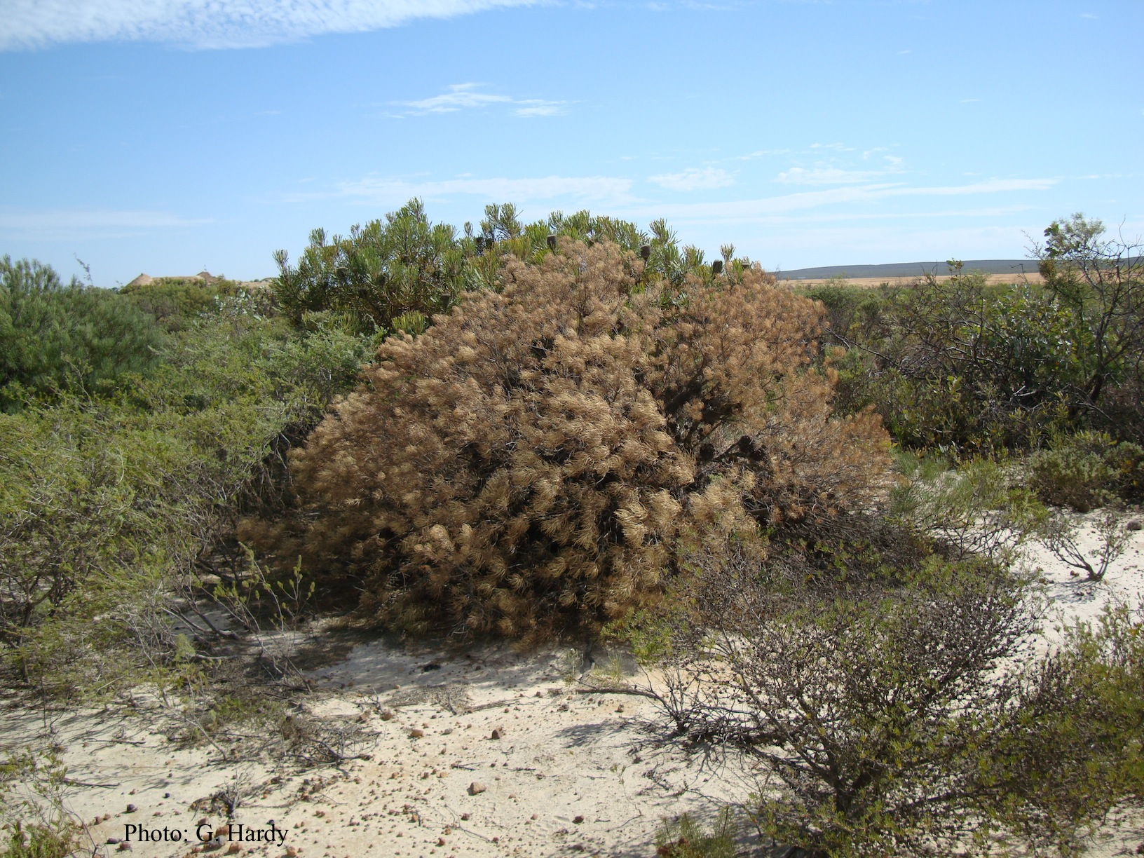 Dead Banksia sp. in a Kwongan heathland on mineral sand near Eneabba, Western Australia recently killed by root and collar rot caused by Phytophthora arenaria |
|
P. ramorum oogonium 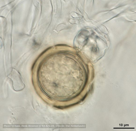 Oogonium with thick oogonial wall, photo from Q-bank, used with permission |
P. katsurae sporangia 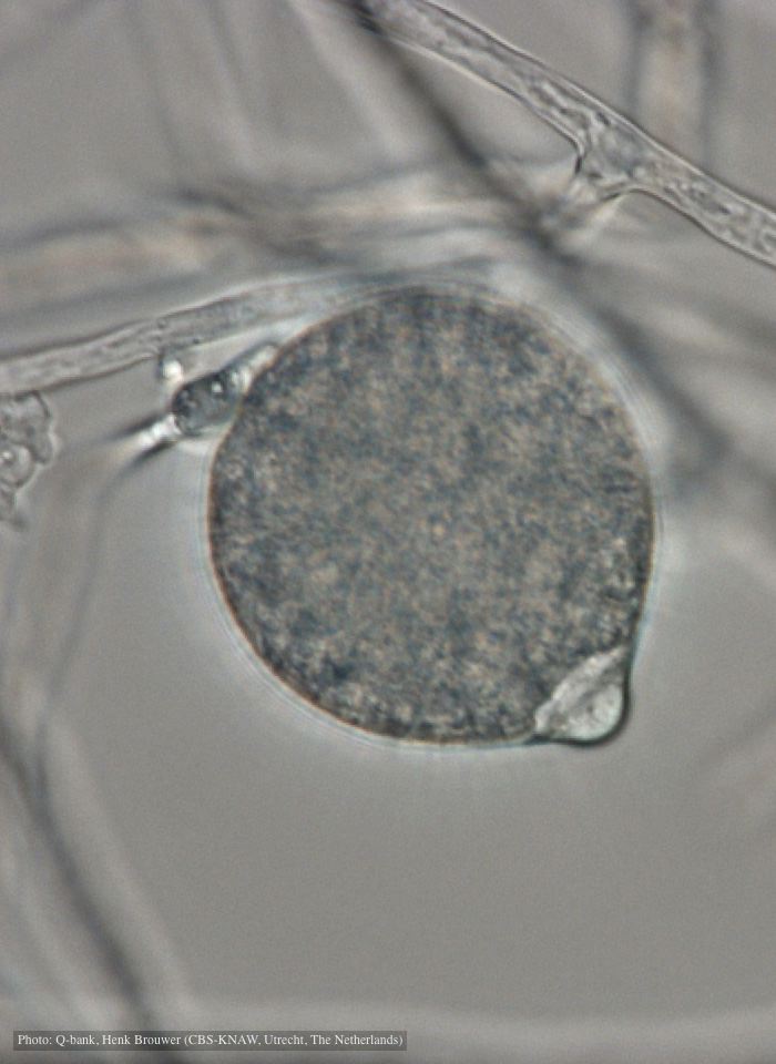 Papillate, non-caducous sporangium with differentiated content; photo used with permission from Q-bank |
P. austrocedrae necrotic lesion in phloem 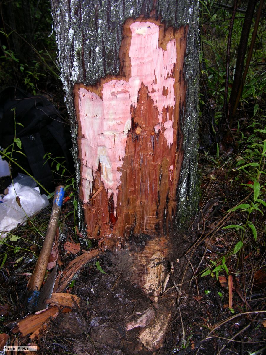 P. austrocedrae - necrotic lesion in phloem |
|
P. megasperma sporangia 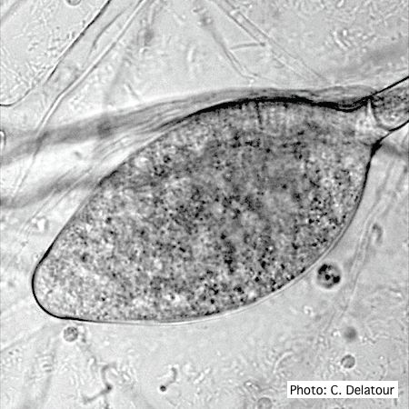 Ovoid, non-papillate sporangia showing internal proliferation of sporangiophore |
P. austrocedrae hyphal swellings in liquid media drawing  Morphology of hyphae of Phytophthora austrocedrae, from Greslebin et al. 2007 |
P. austrocedrae semipapillate sporangium  P. austrocedrae - semipapillate sporangium with off-center attachment. |