

Outplanted California mugwort (Artemisia douglasiana) infected with P. tentaculata, 4.5 years after planting. Plant shows stunting and chlorosis. (P. cryptogea and P. lacustris were also baited from roots/soil of this plant).
Photo Gallery
Site will be retired 9/1/2026
This site is no longer being developed and will be retired on September 1, 2026. Please contact us if you have any questions or would like to provide support to continue the project.
|
P. tentaculata disease symptoms on California mugwort 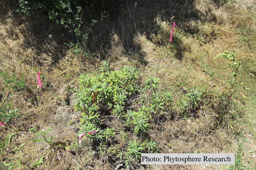 |
P. nemorosa oogonium 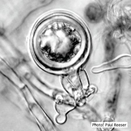 Oogonium with amphigynous antheridium |
P. kernoviae disease on beech 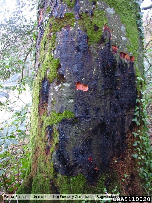 External lesion; 14 November 2003 |
|
Growth of P. arenaria on half-strength PDA 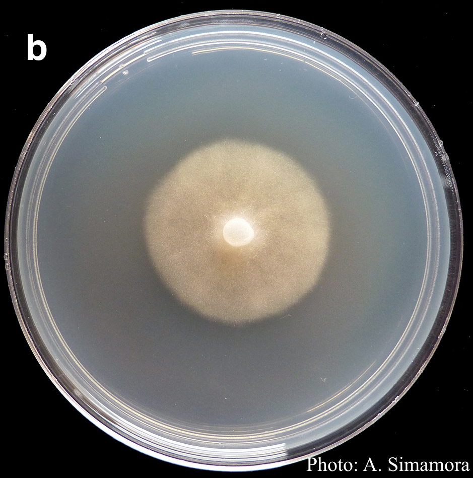 Colony morphology of Phytophthora arenaria after 7 days at 20°C on half-strength PDA |
Comparative gametangial morphology of Phytophthora Clade 5 species  Comparative gametangial morphology of Phytophthora Clade 5 species, with SEM (top) and light microscopy (bottom). P. heveae has smooth walled oogonia with funnel-shaped, amphigynous antheridia. P. agathidicida has mildly stipulate oogonia with globose amphigynous antheridia. P.cocois has mildly bullate oogonia with reflexed amphigynous antheridia. P. castaneae has coarsely bullate oogonium with rugose protuberances and narrow amphigynous antheridia (Weir et al. 2015). |
Phytophthora taxon Agathis bole canker  Canker on a Kauri tree, New Zealand |
|
P. pluvialis on Pinus radiata 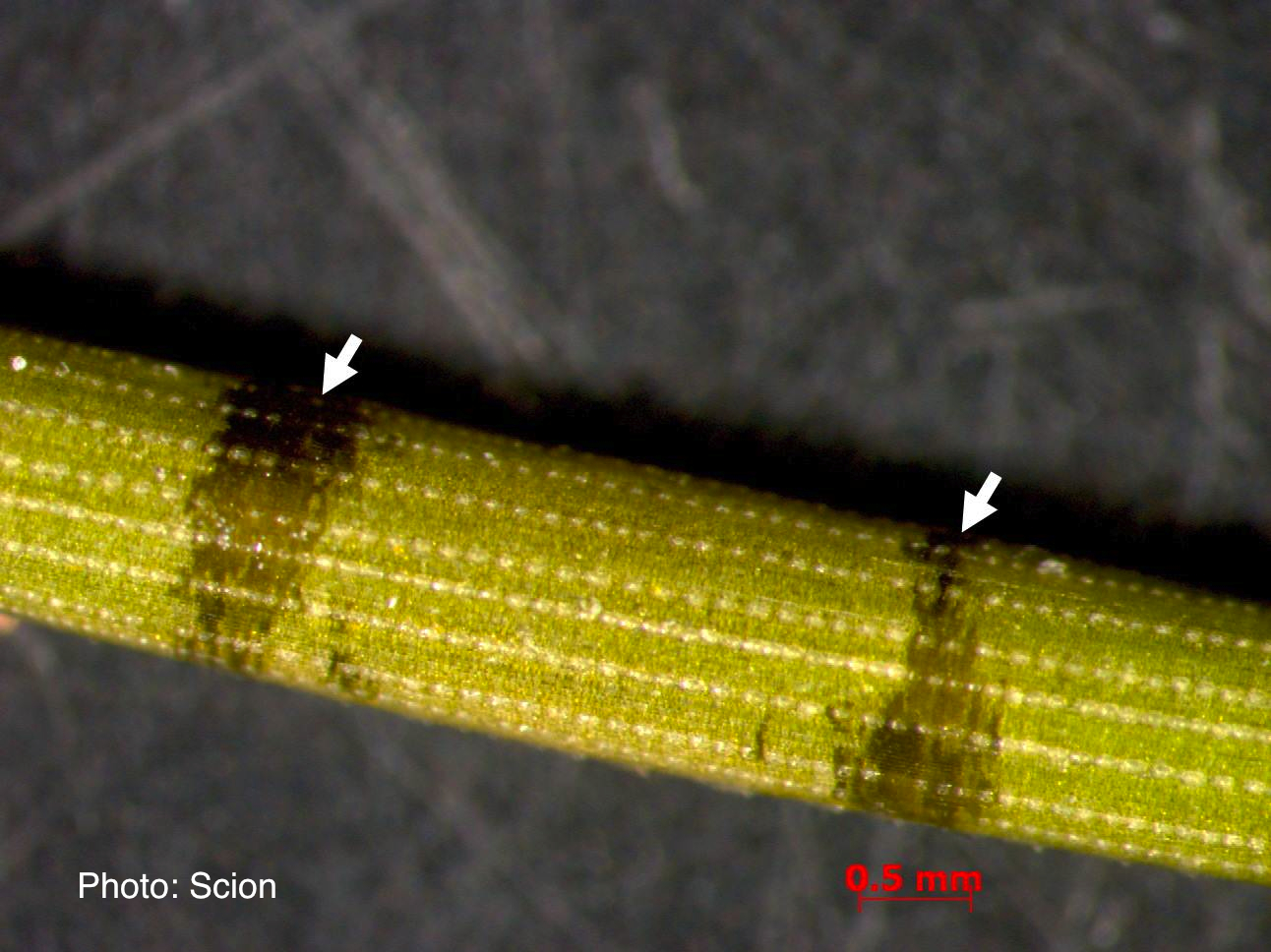 A Pinus radiata needle showing black resinous bands or marks consistent with the presence of red needle cast disease. |
P. pluvialis hyphal swellings 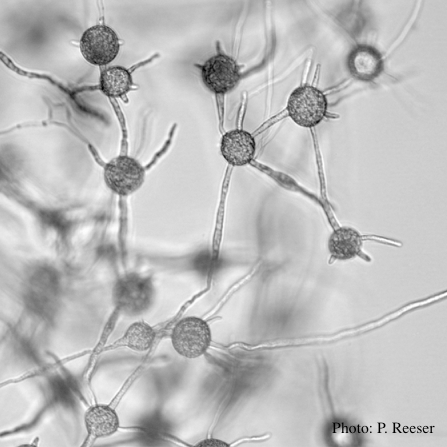 P. pluvialis hyphal swellings in water |
P. kernoviae oogonia 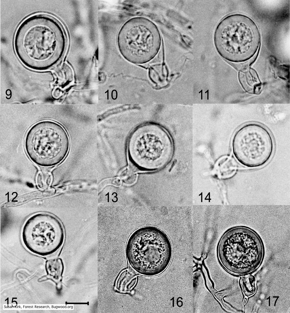 Mycol.Res 109, 853-859; Representative oogonia, antheridia and thick walled plerotic oospores of Phytophthora kernoviae. |
|
P. alni oogonia subspecies and variants 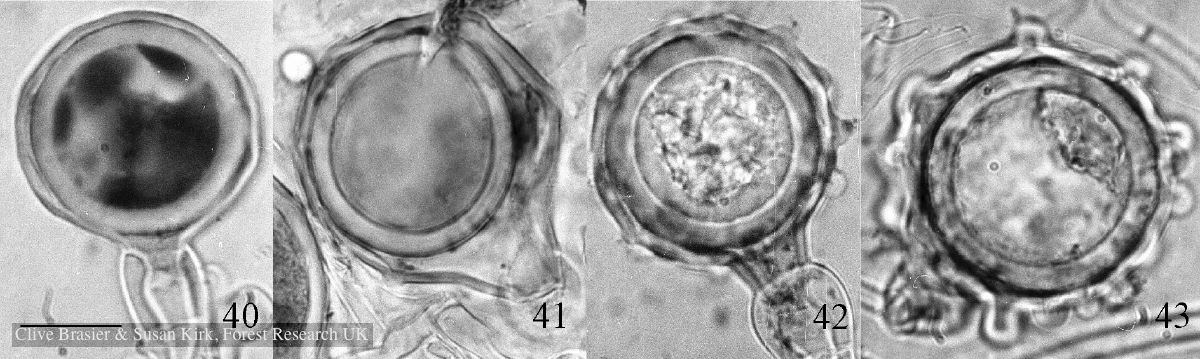 Fig. 40. P. alni subsp. uniformis. Fig. 41. P. alni subsp. multiformis German variant. Fig. 42. P. alni subsp. alni. Fig. 43. |
P. kernoviae sporangium 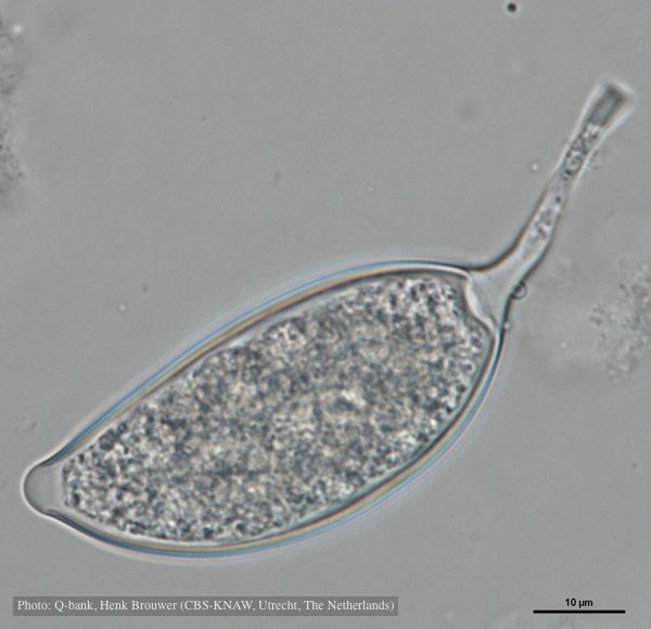 Asymmetrical sporangium, photo from Q-bank, used with permission |
P. pseudosyringae sporangia  Ovoid, semipapillate sporangia showing sympodial development of sporangiophore |