

Colony morphology on PDA at 14 days
Photo Gallery
Site will be retired 9/1/2026
This site is no longer being developed and will be retired on September 1, 2026. Please contact us if you have any questions or would like to provide support to continue the project.
|
P. pseudosyringae colony morphology on PDA  |
P. tentaculata disease symptoms on California mugwort  Nursery grown California mugwort plant (Artemisia douglasiana) infected with P. tentaculata and exhibiting severe root and crown rot |
P. cactorum colony morphology on PDA 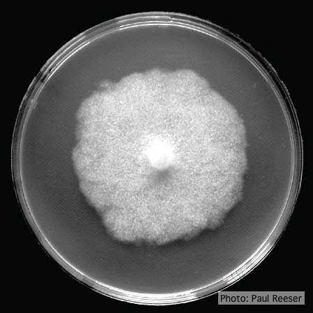 Colony morphology on PDA at 14 days |
|
P. pluvialis on Pinus radiata in New Zealand 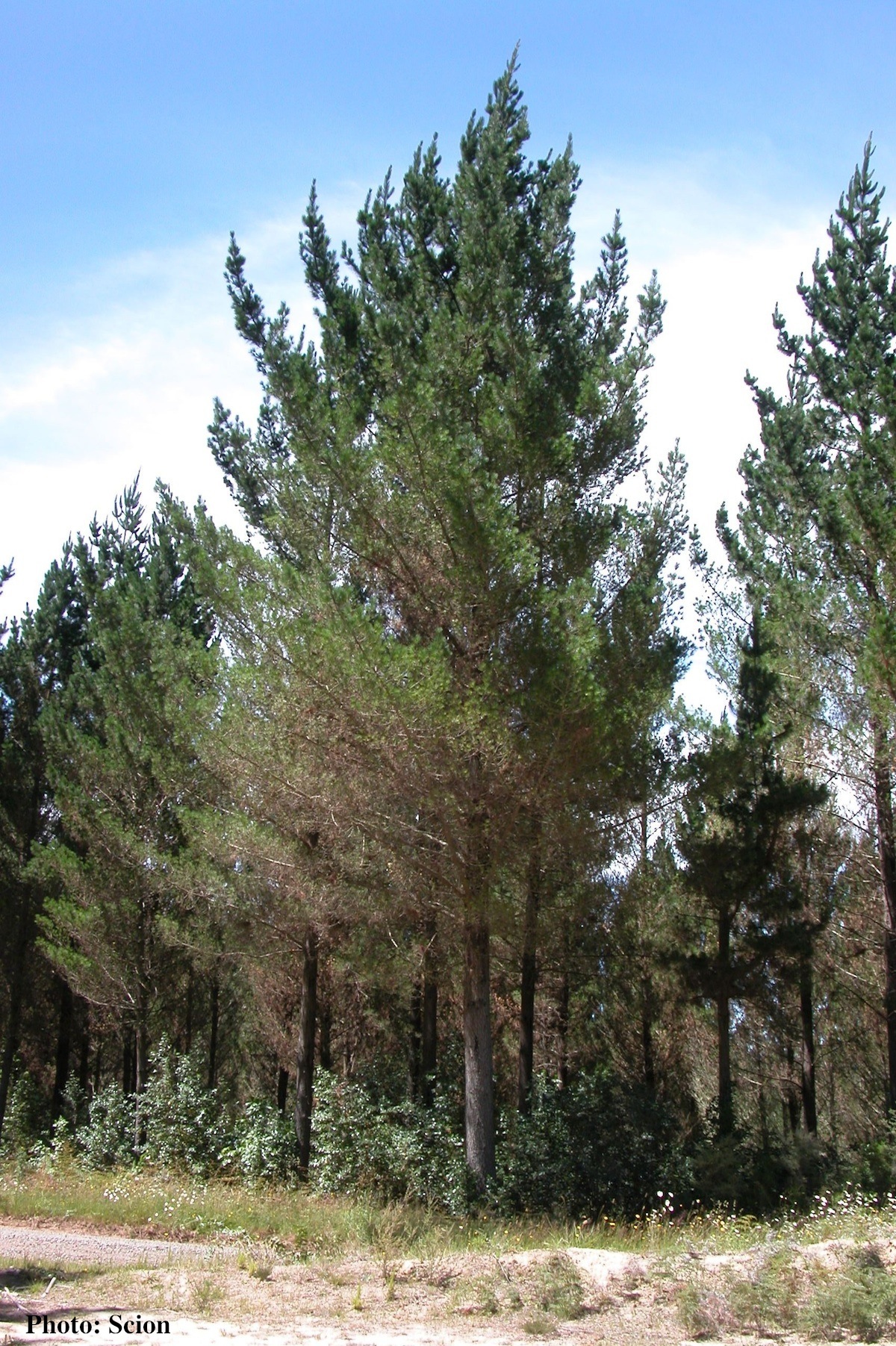 A stand of Pinus radiata trees affected by red needle cast disease. Note that frequently only the lower part of the crown is affected. |
P. cinnamomi hyphal swellings 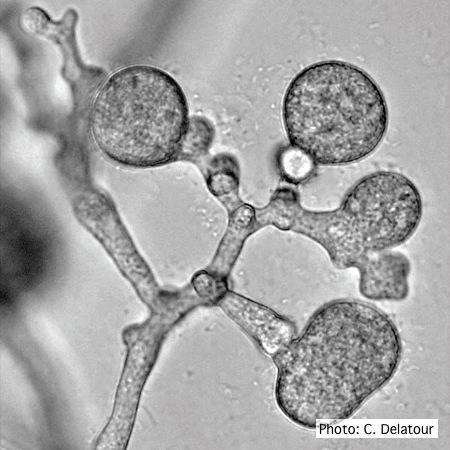 P. cinnamomi hyphal swellings (or thin walled chlamydospores) |
P. cinnamomi on Jarrah 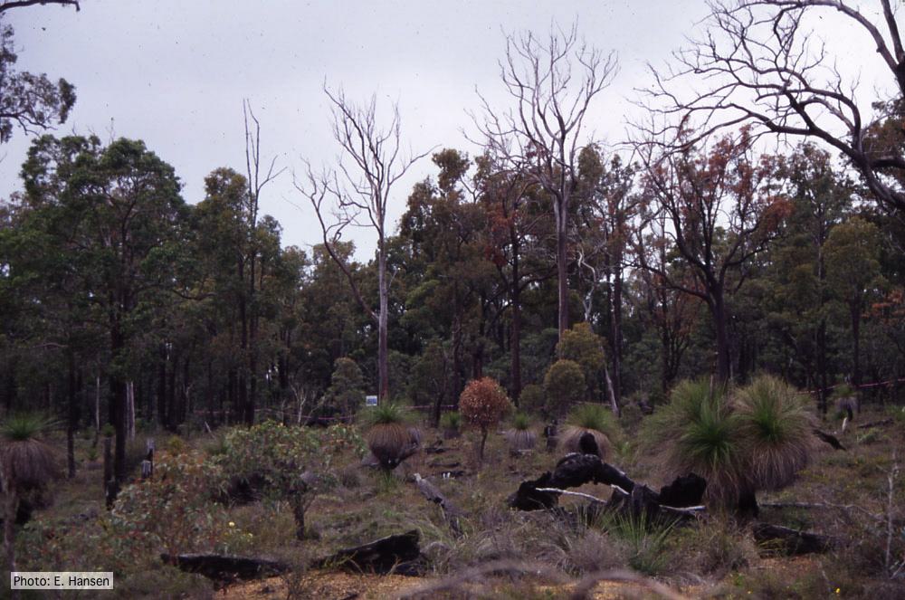 Dieback in Jarrah, Western Australia |
|
P. katsurae growth morphology on β-CMA 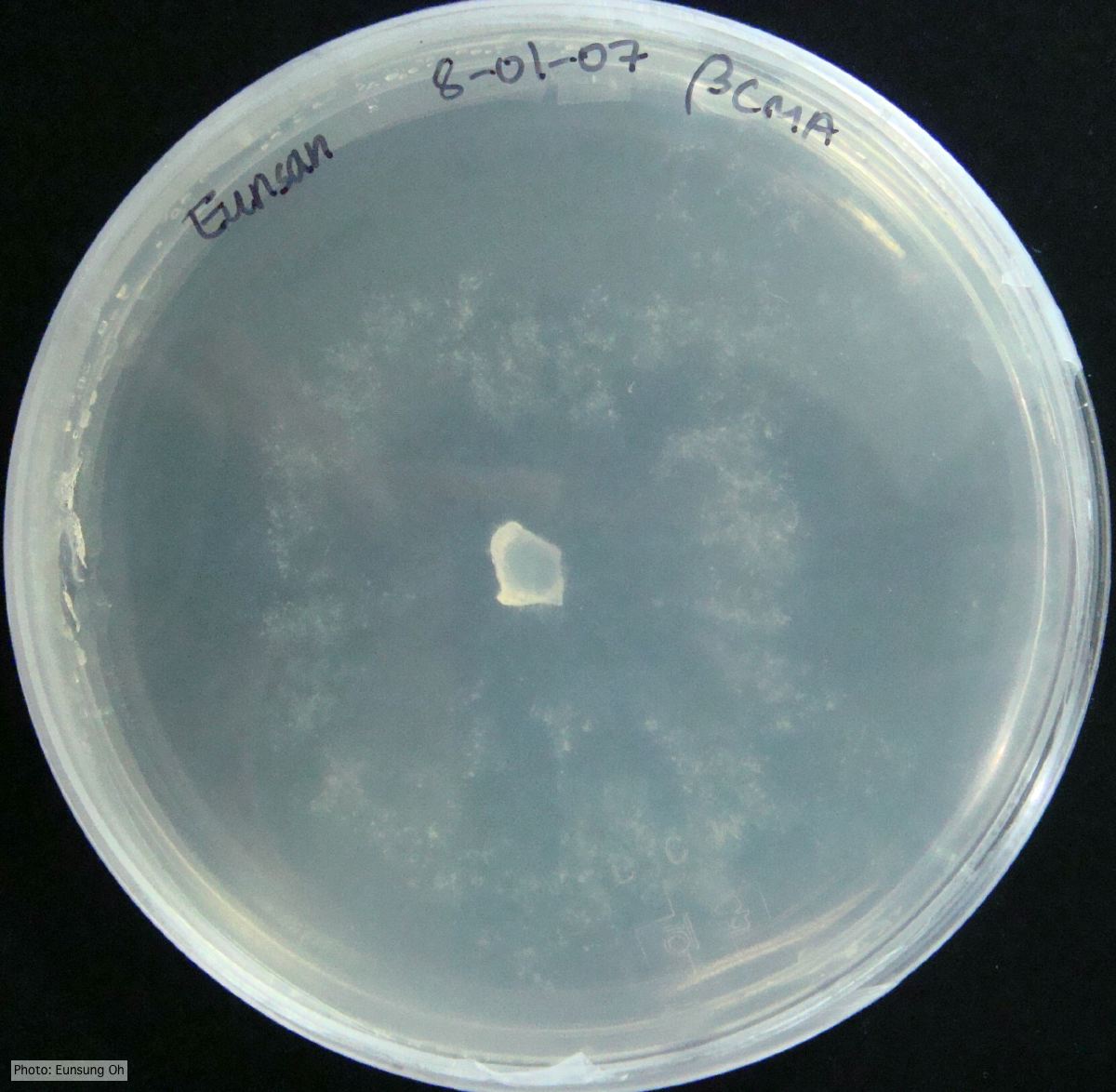 Growth morphology at 7 days on β-CMA |
P. megasperma colony morphology on PDA  Colony morphology on PDA at 7 days |
P. nicotianae symptoms 2  Symptoms of gummosis on black wattle (Fitopatol. bras. 2005) |
|
P. tentaculata oospores and antheridia  Paragynous antheridium attached to oogonium with oospore |
P. lateralis on Port Orford cedar  Dying Chaemacyparis lawsoniana trees in Lopérec, France. |
P. boehmeriae oogonia  Oogonia and oospores with amphigynous antheridia |