

Symptoms of gummosis on black wattle (Fitopatol. bras. 2005)
Photo Gallery
Site will be retired 9/1/2026
This site is no longer being developed and will be retired on September 1, 2026. Please contact us if you have any questions or would like to provide support to continue the project.
|
P. nicotianae symptoms 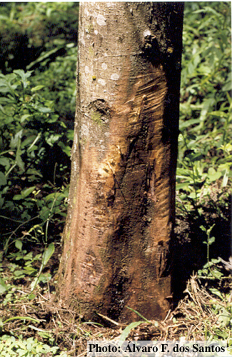 |
P. cambivora inactive lesion on chinquapin  Inactive lesion of P. cambivora on chinquapin |
P. arenaria sporangia 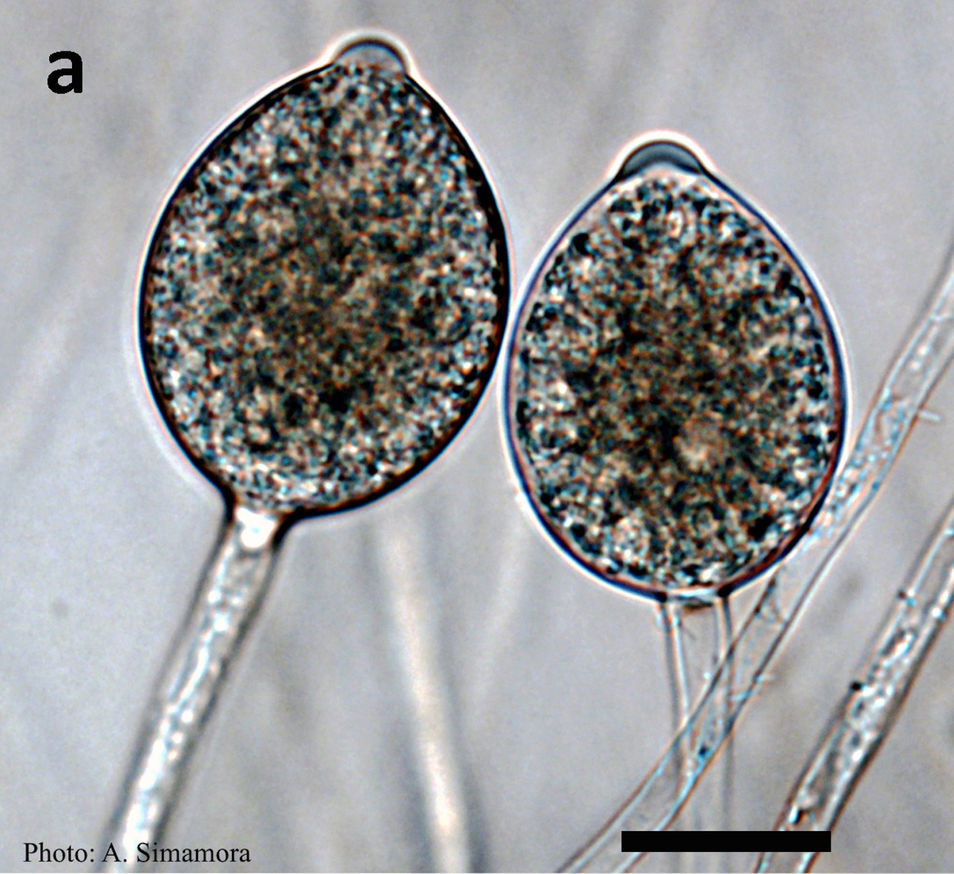 Typical ovoid papillate sporangia of Phytophthora arenaria on V8 agar flooded with soil extract |
|
P. cactorum bleeding canker  Bleeding canker on red oak (Quercus rubra) |
P. austrocedrae - Mal del ciprés in Argentina 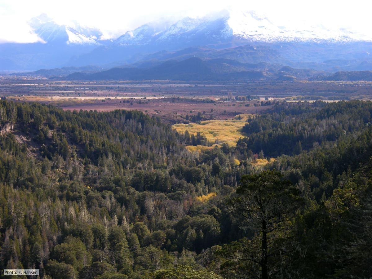 Mal del ciprés looking toward Rio Grande, Chubut Province, Argentina |
P. agathidicia growth on PDA 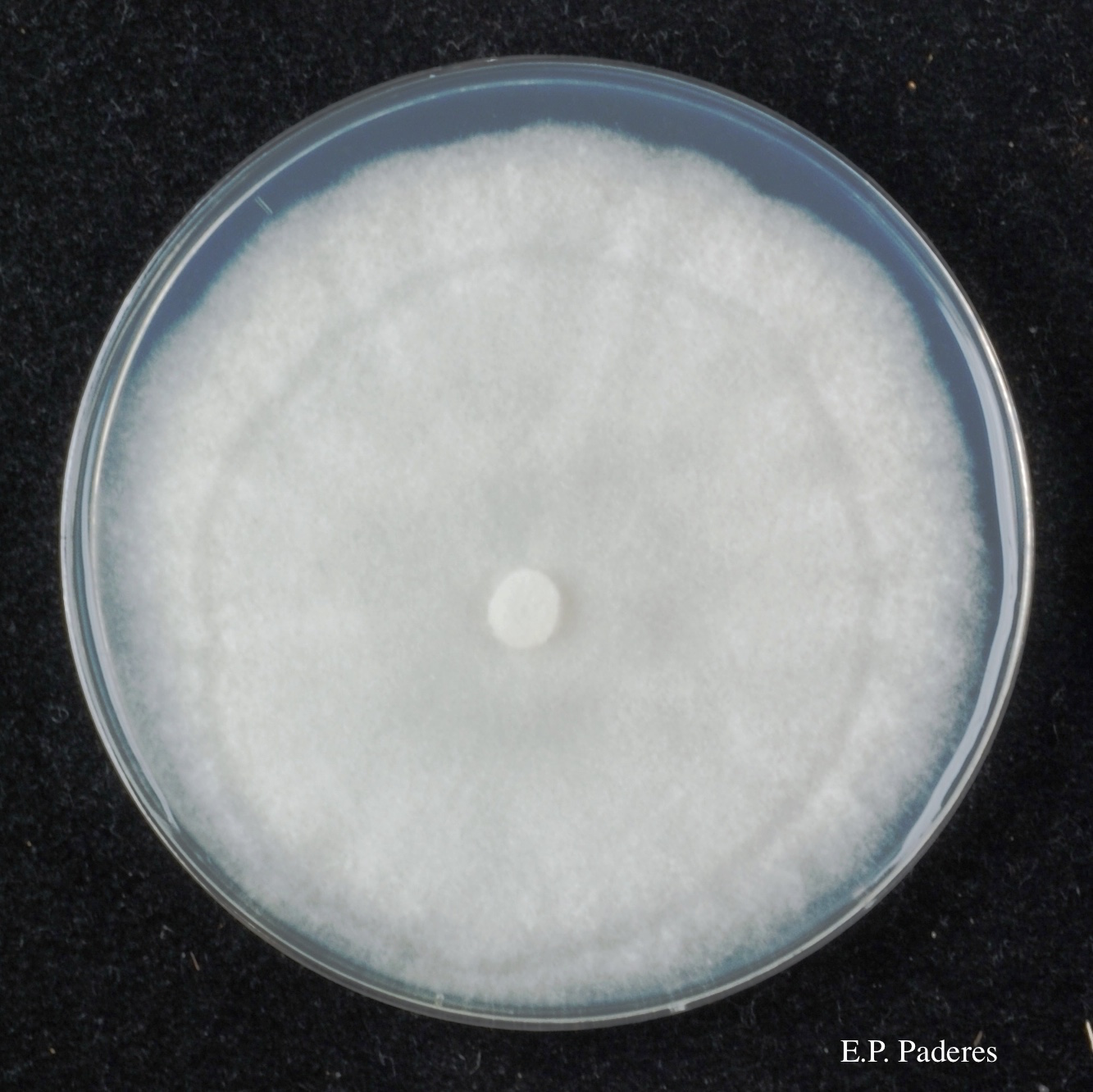 Colony morphology of ex-holotype ICMP 17027 after 10-days incubation at 20°C in the dark |
|
Comparative gametangial morphology of Phytophthora Clade 5 species  Comparative gametangial morphology of Phytophthora Clade 5 species, with SEM (top) and light microscopy (bottom). P. heveae has smooth walled oogonia with funnel-shaped, amphigynous antheridia. P. agathidicida has mildly stipulate oogonia with globose amphigynous antheridia. P.cocois has mildly bullate oogonia with reflexed amphigynous antheridia. P. castaneae has coarsely bullate oogonium with rugose protuberances and narrow amphigynous antheridia (Weir et al. 2015). |
P. chlamydospora chlamydospore 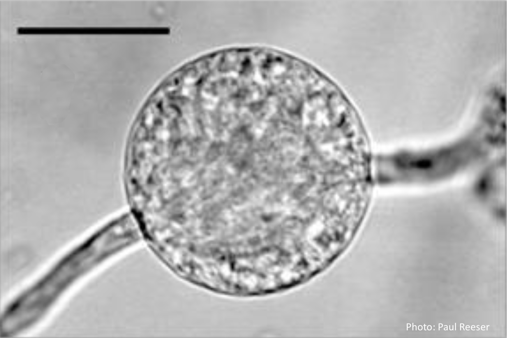 Phytophthora chlamydospora chlamydospore in agar. Bar is 20µm. |
P. tentaculata microscopic characteristics 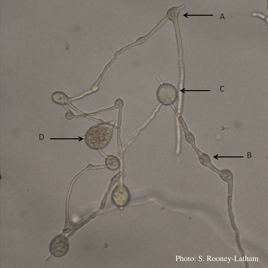 Hyphal swellings occuring at branching points of Mycelium (A), Intercalary hyphal swellings (B), Chlamydospore (C ), Sporangia (D) |
|
P. nicotianae colony morphology on CMA  Phytophthora nicotianae CBS 321.49 CMA after 7 days at 24 degrees. Photo from Q-bank: www.q-bank.eu, Henk Brouwer (CBS-KNAW, Utrecht, The Netherlands) |
P. pseudotsugae sporangia  Broadly ovoid, papillate sporangia in water |
P. cactorum oogonium 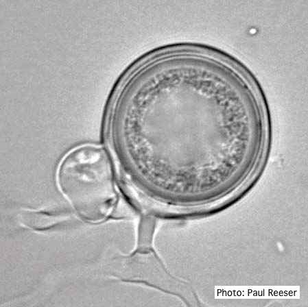 Oogonium with paragynous antheridia close to oogonial stalk. Oospores are slightly aplerotic. |