

A stand of Pinus radiata trees affected by red needle cast disease. Note the reddish appearance of affected trees prior to needle drop.
Photo Gallery
Site will be retired 9/1/2026
This site is no longer being developed and will be retired on September 1, 2026. Please contact us if you have any questions or would like to provide support to continue the project.
|
P. pluvialis on Pinus radiata in New Zealand 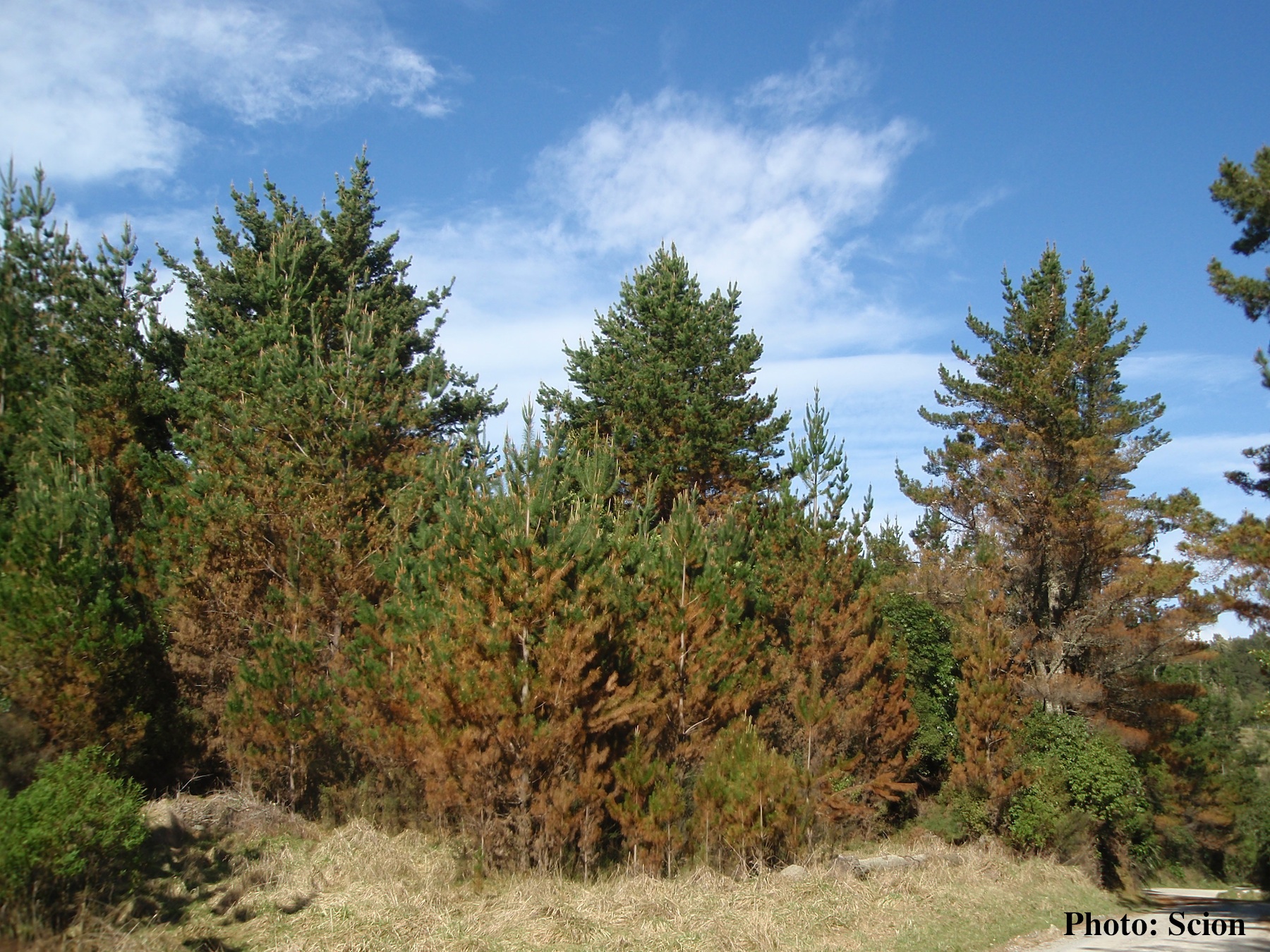 |
P. pluvialis symptoms on Douglas-fir  Red needle cast symptoms on Douglas-fir in western Oregon, 2015 |
Stain from Port Orford Cedar root disease  Stain from Chamaecyparis lawsoniana root disease on the Smith River |
|
P. pluvialis sporangium 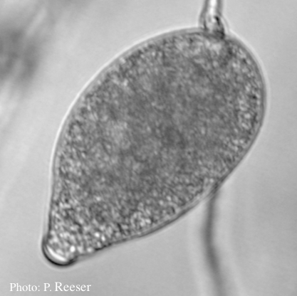 Sporangium showing typical ovoid shape and semi-papillate condition |
P. austrocedrae irregular sporangium 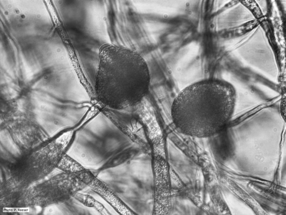 P. austrocedrae - irregular sporangium with lateral attachment and swelling in sporangiophore |
P. megasperma oogonium 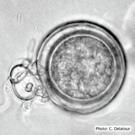 Oogonium with paragynous antheridia applied close to the ogonial stalk. |
|
P. austrocedrae - Mal del ciprés, stages of decline  Mal del ciprés, stages of decline |
P. pinifolia hyphal swellings  Spherical hyphal swelling with radiating hyphae (from Duran et al. 2008). Scale bar = 20 μm. |
P. kernoviae sporangium 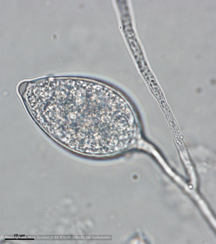 Papillate and caducous sporangium, photo from Q-bank, used with permission |
|
P. lateralis colony morphology on PDA 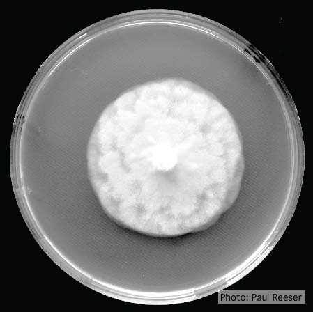 Growth morphology on PDA of Phytophthora lateralis |
P. pseudotsugae amphigynous oogonium 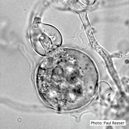 P. pseudotsugae oogonium with amphigynous antheridia |
P. cambivora sporangium with sympodial proliferation 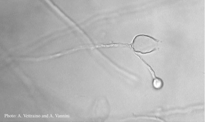 Empty sporagium showing sympodial proliferation |