

Rosaceous colony morphology at 14 days at 20°C on PDA
Photo Gallery
Site will be retired 9/1/2026
This site is no longer being developed and will be retired on September 1, 2026. Please contact us if you have any questions or would like to provide support to continue the project.
|
P. cambivora colony morphology on PDA  |
P. cinnamomi on Banksia 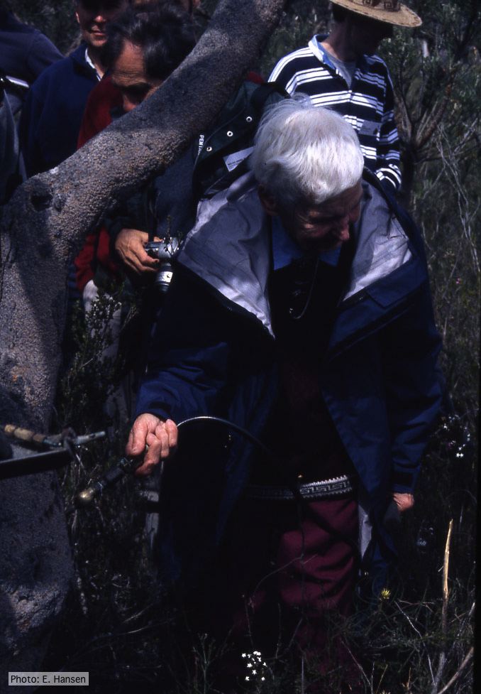 Gretna Weste injecting Banksia with phosphonate |
P. pluvialis sporangia.  P. pluvialis sporangia on tape peel from infected Douglas-fir needle. |
|
Tāne Mahuta “Lord of the Forest” kauri tree 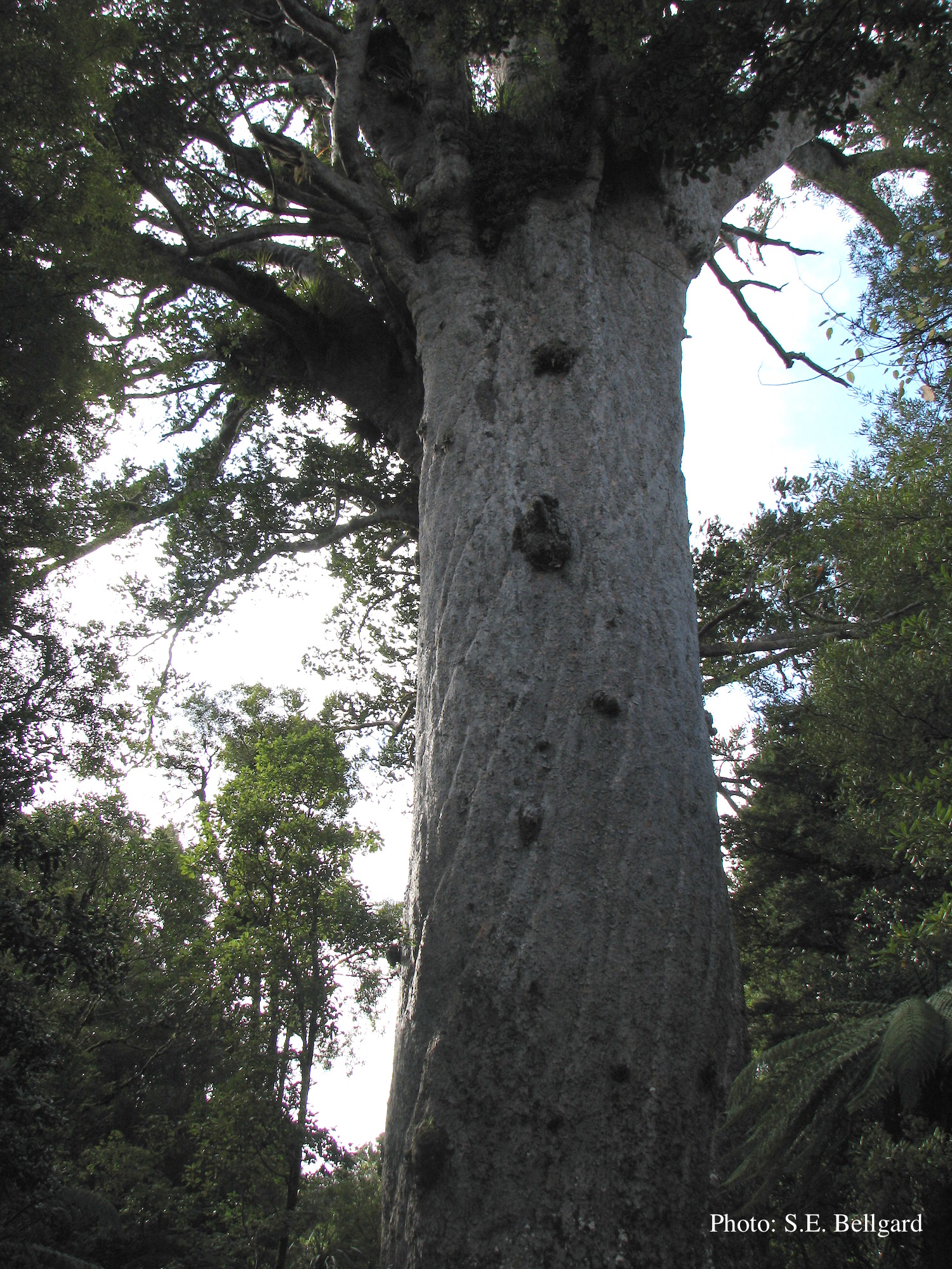 Tāne Mahuta “Lord of the Forest” is a giant kauri tree (approximately 47 metres in height) in the Waipoua Forest of Northland Region, New Zealand. Its age is unknown but is estimated to be between 1,250 and 2,500 years |
P. kernoviae canker  Bole lesion on Fagus sylvatica |
P. pluvialis hyphal swellings 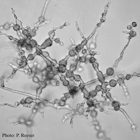 P. pluvialis hyphal swellings on agar |
|
Growth morphology on V8 of P. lateralis 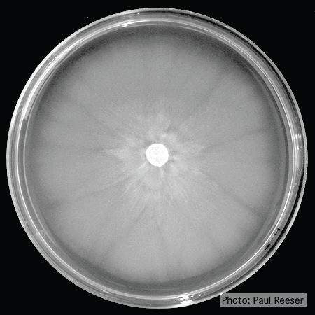 Colony morphology on V8 at 14 days |
P. alni in riparian alder, Scotland 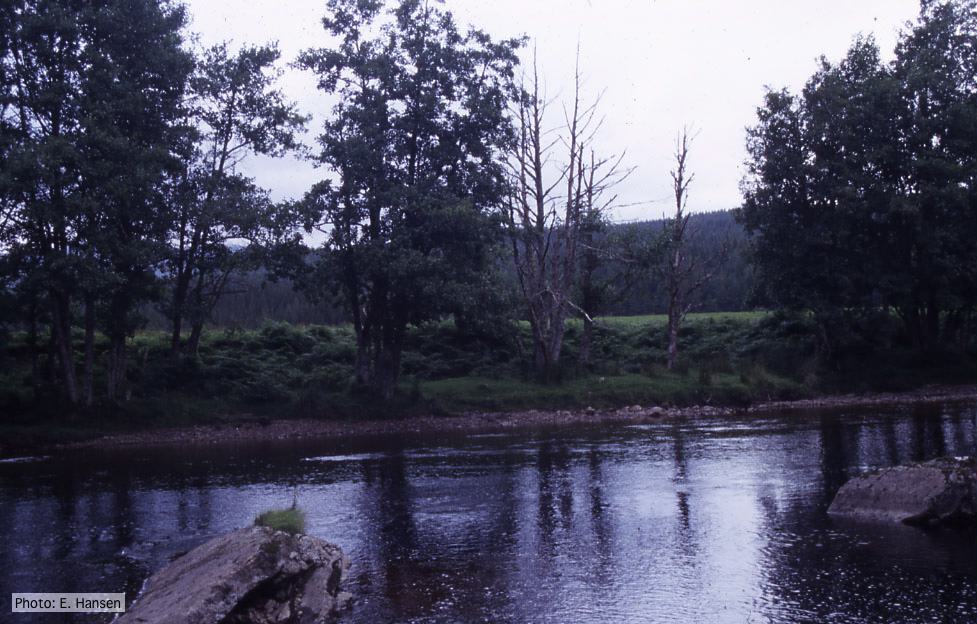 P. alni in riparian alder, Scotland |
P. cambivora sporangium  Ovoid non-papillate sporangia with well-rounded base |
|
P. austrocedrae - Mal del ciprés in Argentina 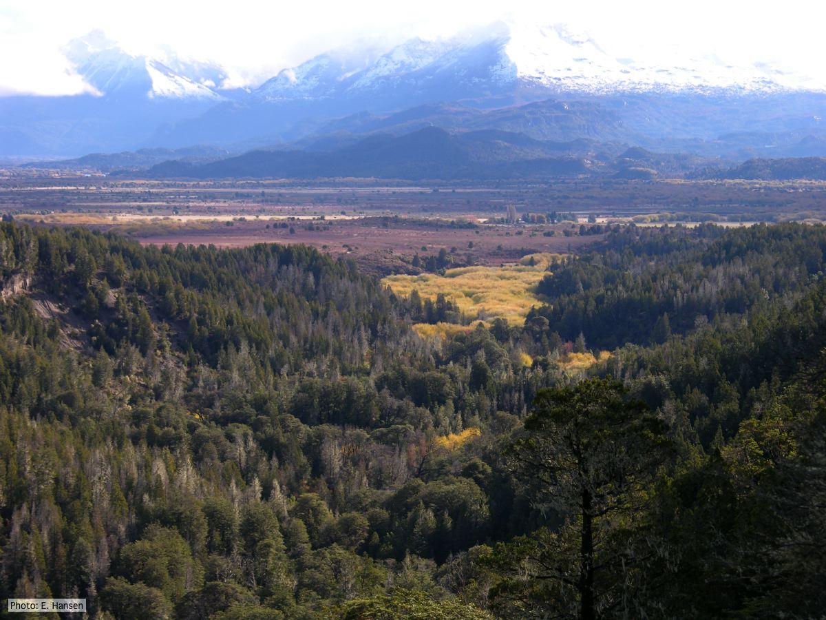 Mal del ciprés looking toward Rio Grande, Chubut Province, Argentina |
P. ramorum colony morphology on V8 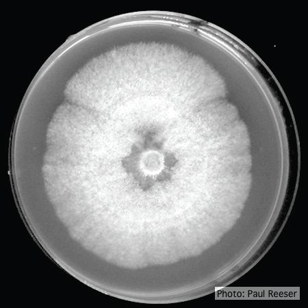 P. ramorum colony morphology on V8 |
P. austrocedrae - Mal del ciprés, stages of decline  Mal del ciprés, stages of decline |