

Colony morphology on V8 at 14 days
Photo Gallery
Site will be retired 9/1/2026
This site is no longer being developed and will be retired on September 1, 2026. Please contact us if you have any questions or would like to provide support to continue the project.
|
P. cambivora colony morphology on V8 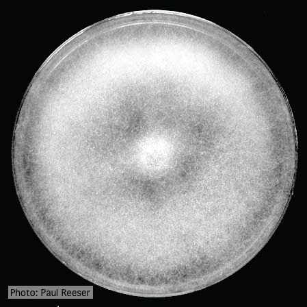 |
P. megakarya sporangia (photo from Q-bank, used with permission). 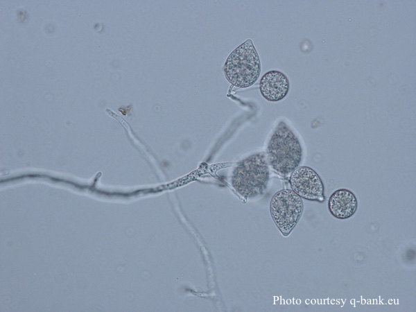 P. megakarya sporangia |
P. tentaculata disease symptoms on sticky monkey flower 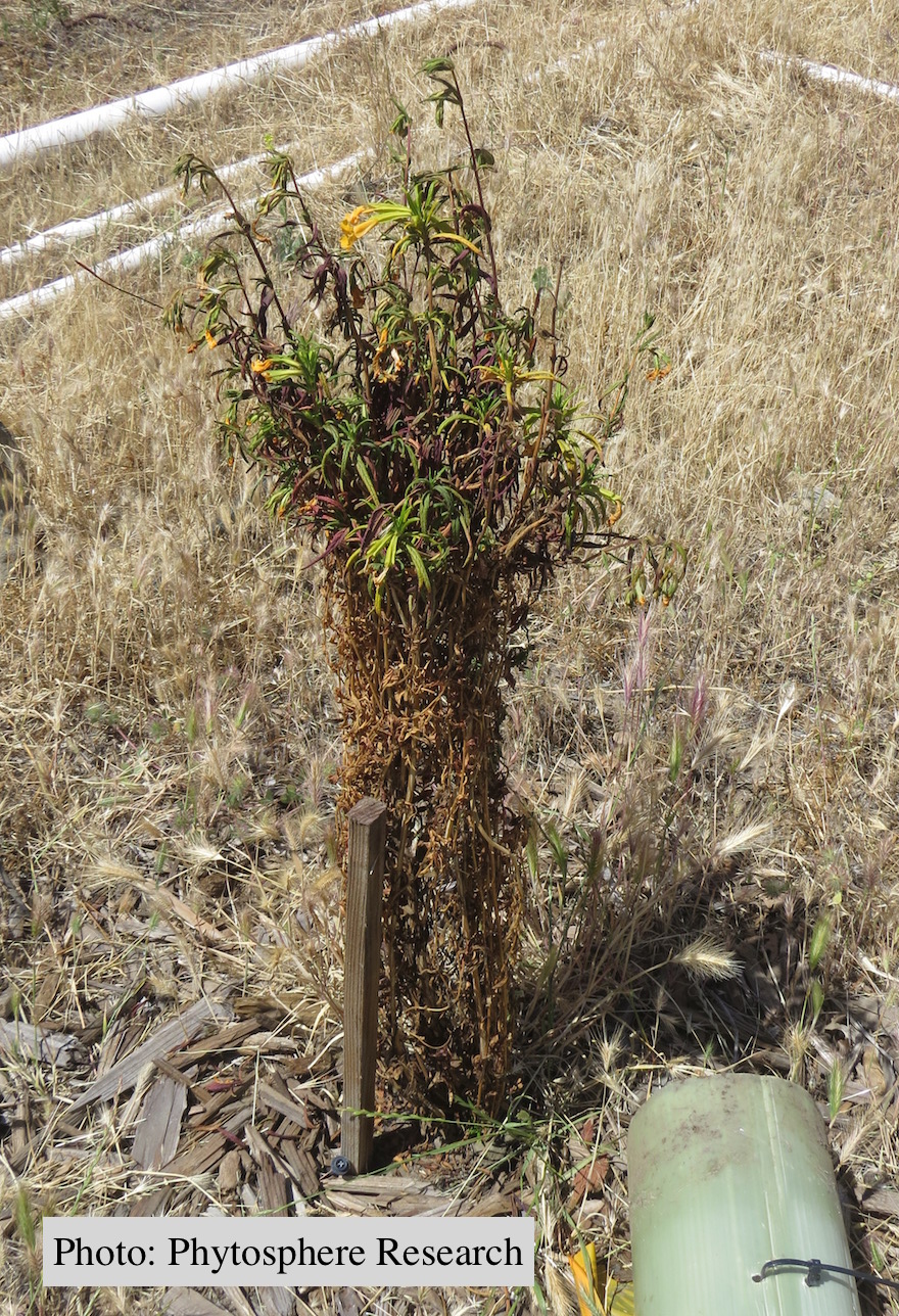 Outplanted sticky monkey flower (Diplacus aurantiacus) infected with P. |
|
P. nicotianae colony morphology on PDA 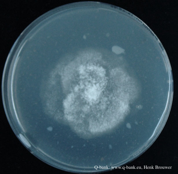 Phytophthora nicotianae CBS 321.49 PDA after 7 days at 24 degrees. Photo from Q-bank: www.q-bank.eu, Henk Brouwer (CBS-KNAW, Utrecht, The Netherlands) |
P. nicotianae oogonia 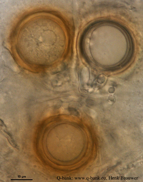 P. nicotianae oogonia 100x. Photo from Q-bank: www.q-bank.eu, Henk Brouwer (CBS-KNAW, Utrecht, The Netherlands) |
P. cinnamomi in Australia  Death of Xanthoria and other native vegetation, Victoria, Australia |
|
P. pseudosyringae sporangia  Ovoid, semipapillate sporangia showing sympodial development of sporangiophore |
P. pseudotsugae amphigynous oogonium 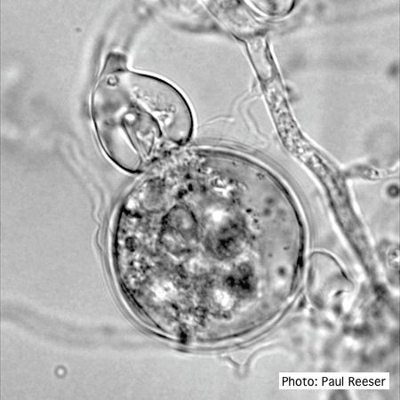 P. pseudotsugae oogonium with amphigynous antheridia |
P. alni in riparian alder, Scotland 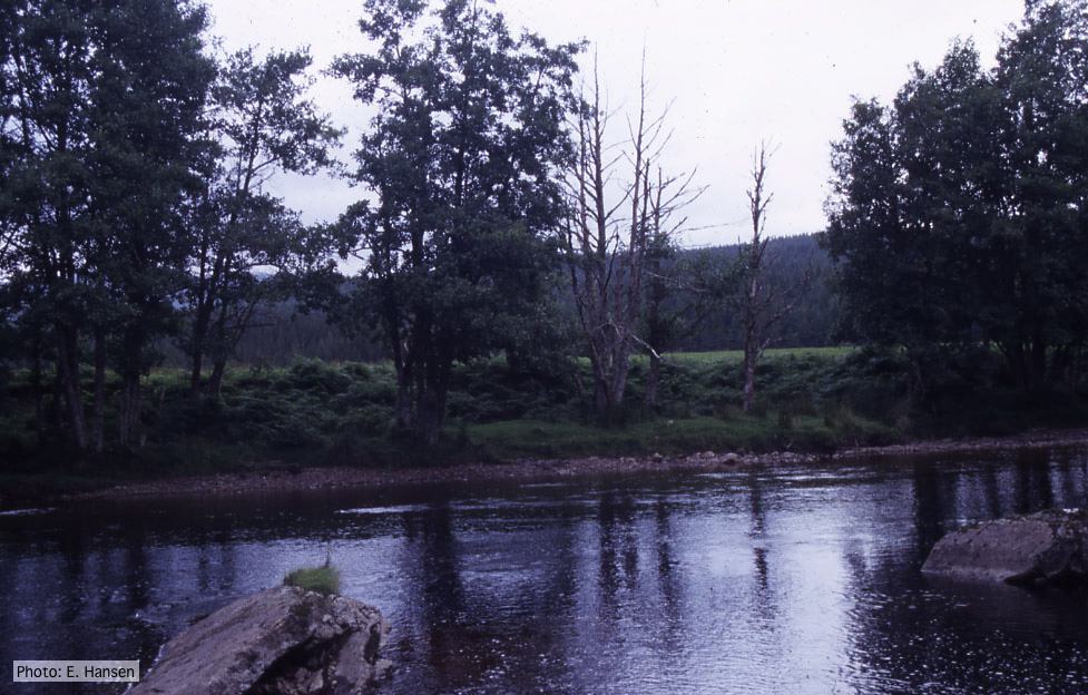 P. alni in riparian alder, Scotland |
|
P. pluvialis colony morphology on carrot agar  Colony morphology on carrot agar at 20 days |
P. cambivora sporangium with sympodial proliferation 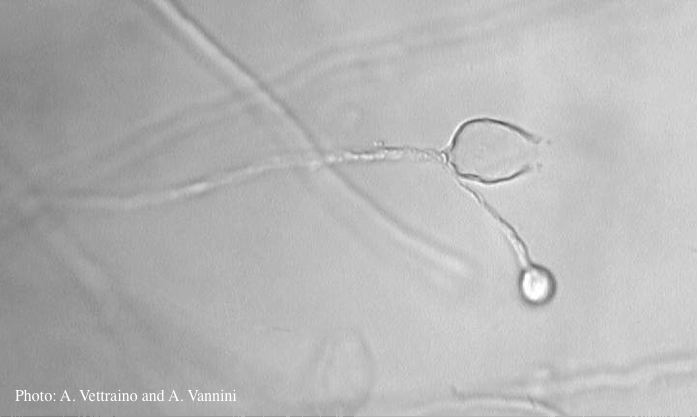 Empty sporagium showing sympodial proliferation |
P. pinifolia coenocytic hyphae 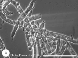 Coenocytic hyphae (from Duran et al. 2008). Scale bar = 20 μm. |