

Colony morphology of Phytophthora arenaria after 7 days at 20°C on V8 agar
Photo Gallery
|
Growth of P. arenaria on V8  |
P. pinifolia on Pinus radiata  Pinus radiata, note Infected needles at right angles to stem |
P. ramorum leaf symptoms on tan oak 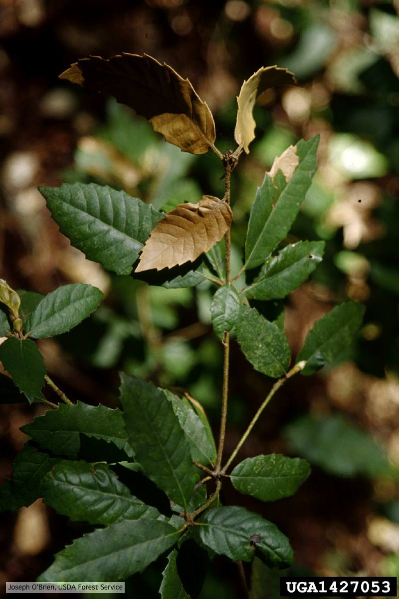 Tip symptoms on tanoak seedling (Notholithocarpus densiflorus). |
|
P. tentaculata sporangium  Papillate sporangium of P. tentaculata |
P. pluvialis sporangium 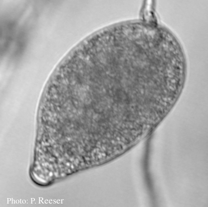 Sporangium showing typical ovoid shape and semi-papillate condition |
P. megasperma sporangia 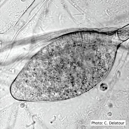 Ovoid, non-papillate sporangia showing internal proliferation of sporangiophore |
|
P. lateralis on Port Orford cedar  Atypical decline caused by aerial infections in Scaër, France |
P. frigida sporangia  Noncaducous sporangia showing ovoid shape and papillate condition |
Chlamydospore of P. lateralis 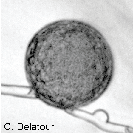 Laterally intercalary chlamydospore of Phytophthora lateralis |
|
Necrotic lesion in phloem caused by P. austrocedrae  Necrotic lesion in phloem with resin pocket caused by P. austrocedrae |
P. siskiyouensis canker on Italian alder 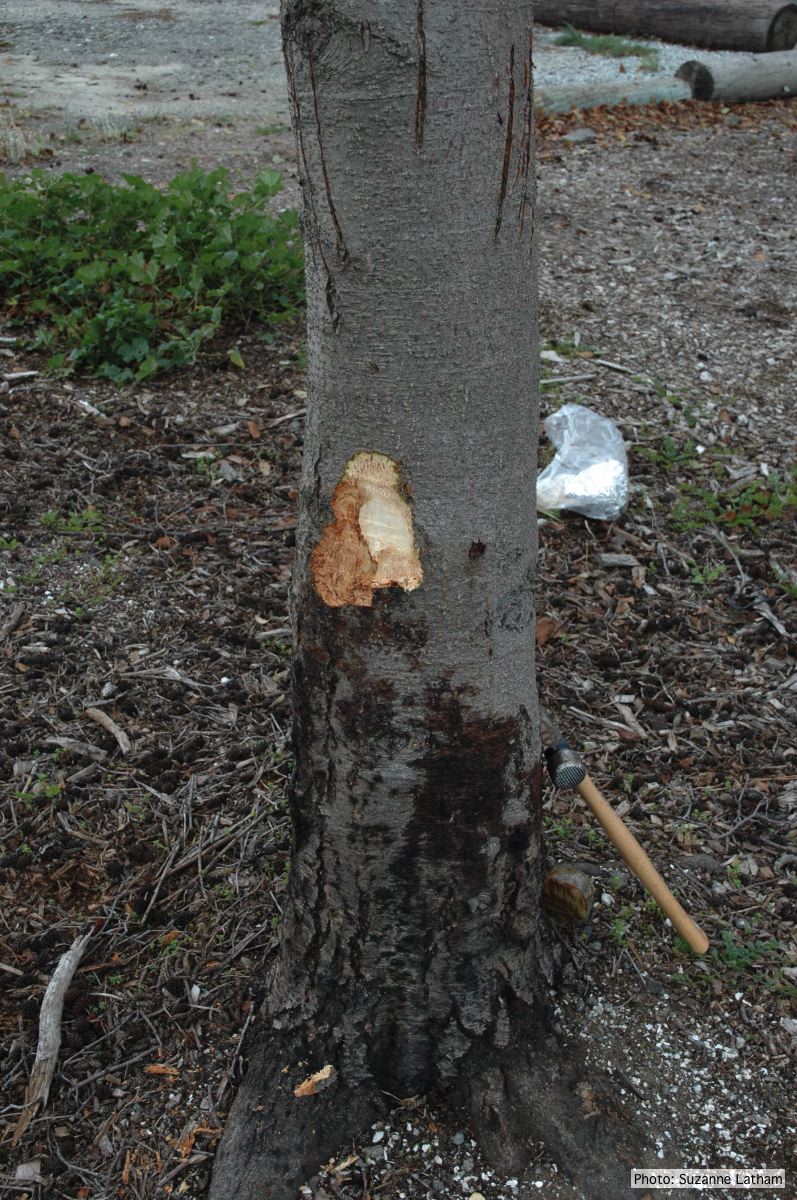 Bole lesions in the tissues under the bark of a bleeding canker: distinct margin between healthy and disease tissues |
P. agathadicida disease symptom 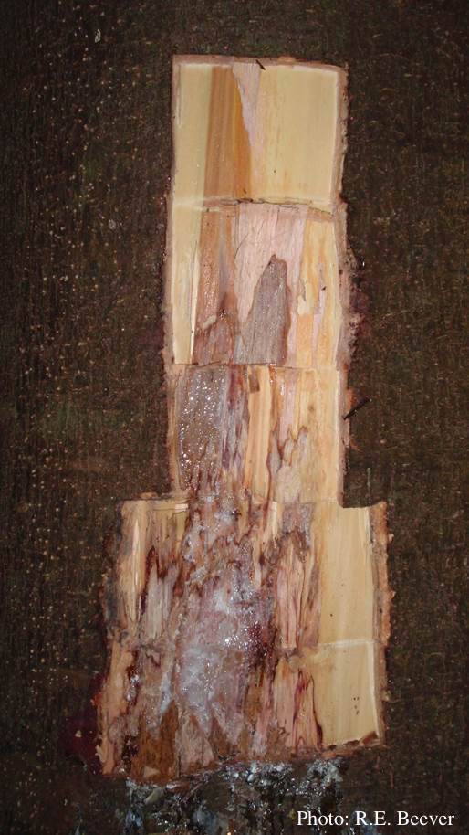 Excavated lesion, with outer bark removed showing extent of disease-front |