

Colony morphology of P. pinifolia at 20°C on CMA-NARP after 3 weeks. From Duran et al. 2008
Photo Gallery
|
P. pinifolia colony morphology on CMA-NARP 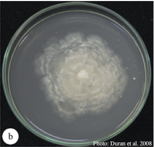 |
P. cryptogea sporangium  Ovoid non-papillate sporangia in water. |
P. tentaculata disease symptoms on California mugwort 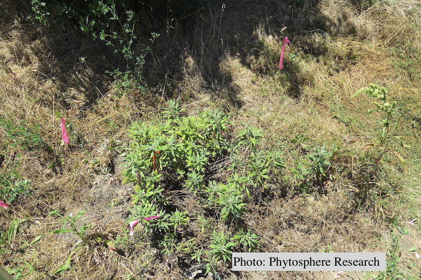 Outplanted California mugwort (Artemisia douglasiana) infected with P. tentaculata, 4.5 years after planting. Plant shows stunting and chlorosis. (P. cryptogea and P. lacustris were also baited from roots/soil of this plant). |
|
P. kernoviae sporangium 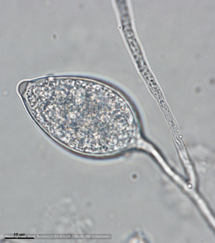 Papillate and caducous sporangium, photo from Q-bank, used with permission |
P. kernoviae canker  Bole lesion on Fagus sylvatica |
P. pluvialis - appearance of new growth 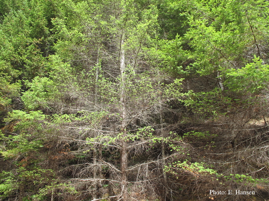 Tufted appearance of new growth from surviving buds on Douglas-fir, one year after defoliation. |
|
P. kernoviae colony morphology on V8 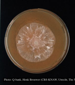 Colony morphology at 7 days at 18°C on V8, photo from Q-bank, used with permission. |
P. megakarya sporangia (photo from Q-bank, used with permission). 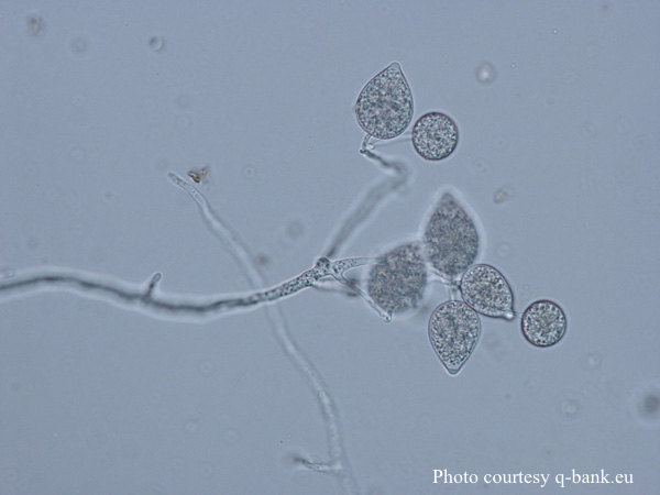 P. megakarya sporangia |
P. ramorum sporangium 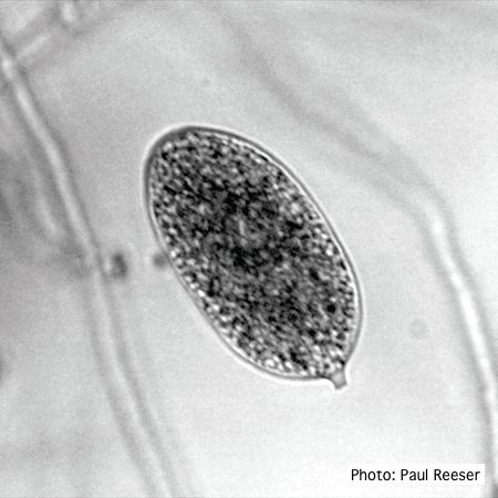 P. ramorum sporangium |
|
Growth morphology on V8 of P. lateralis 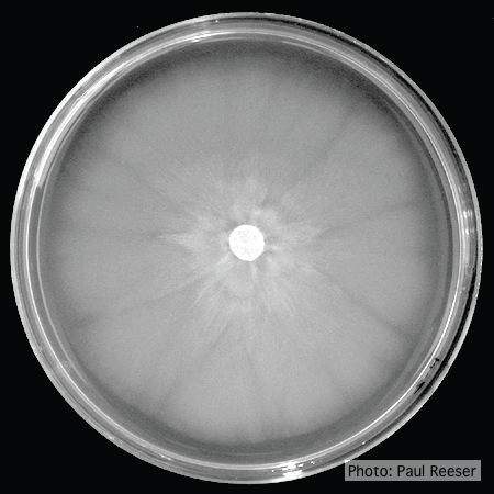 Colony morphology on V8 at 14 days |
P. cactorum bleeding canker 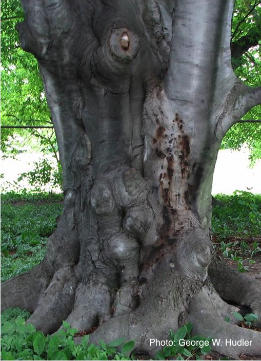 Bleeding canker on European beech (Fagus sylvatica) |
P. pinifolia on Pinus radiata 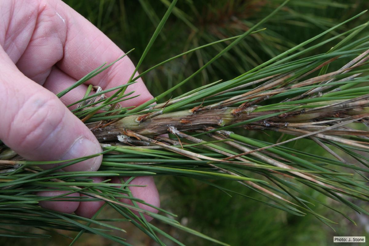 Pinus radiata needles, note “black line” symptom near needle bases |