

Hyphal swellings occuring at branching points of Mycelium (A), Intercalary hyphal swellings (B), Chlamydospore (C ), Sporangia (D)
Photo Gallery
|
P. tentaculata microscopic characteristics 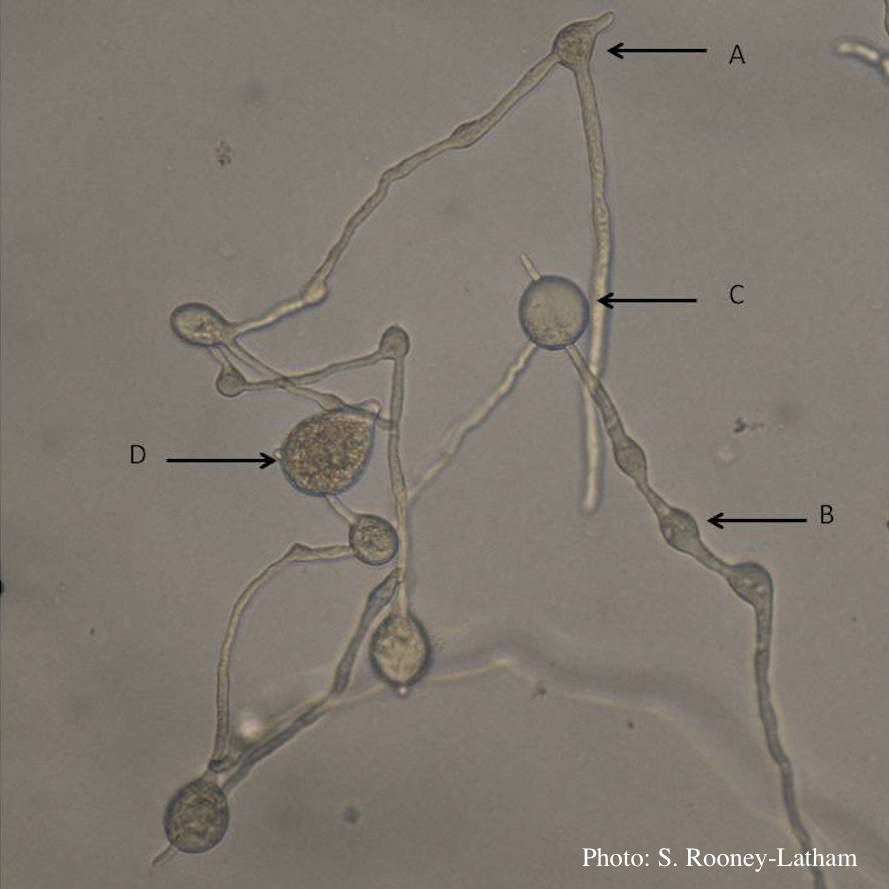 |
P. cambivora colony morphology on MA  Appressed colony morphology at 14 days at 20°C on MA |
Growth of P. arenaria on MEA 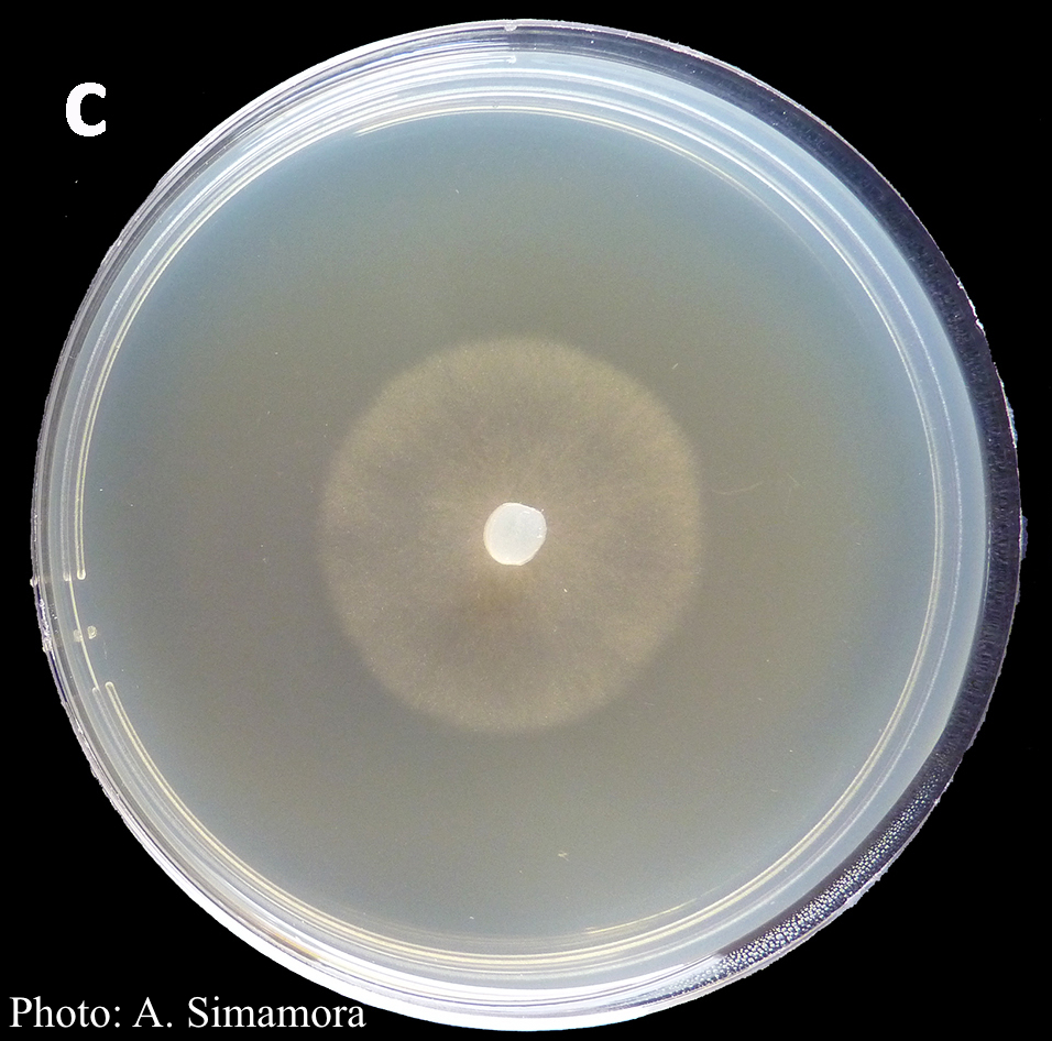 Colony morphology of Phytophthora arenaria after 7 days at 20°C on malt extract agar |
|
P. ramorum sporangia and chlamydospores 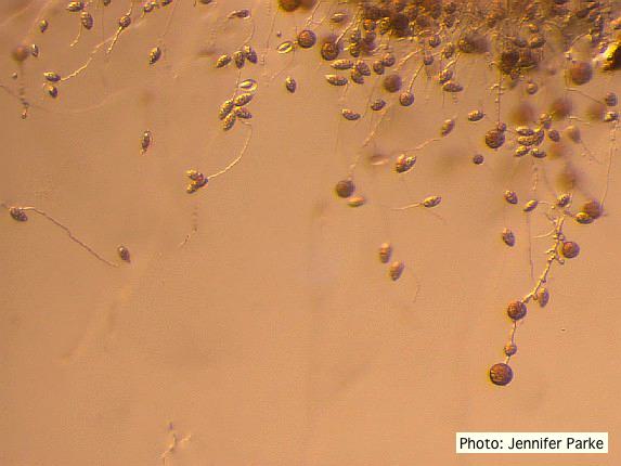 Sporangia and chlamydospores of P. ramorum |
P. kernoviae sporangium 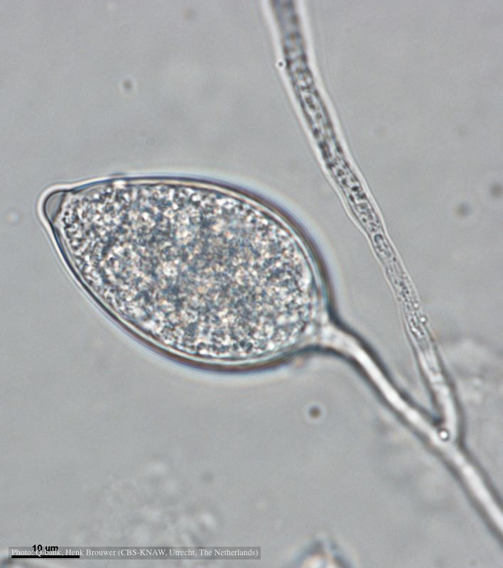 Papillate and caducous sporangium, photo from Q-bank, used with permission |
Healthy Port Orford Cedar tree 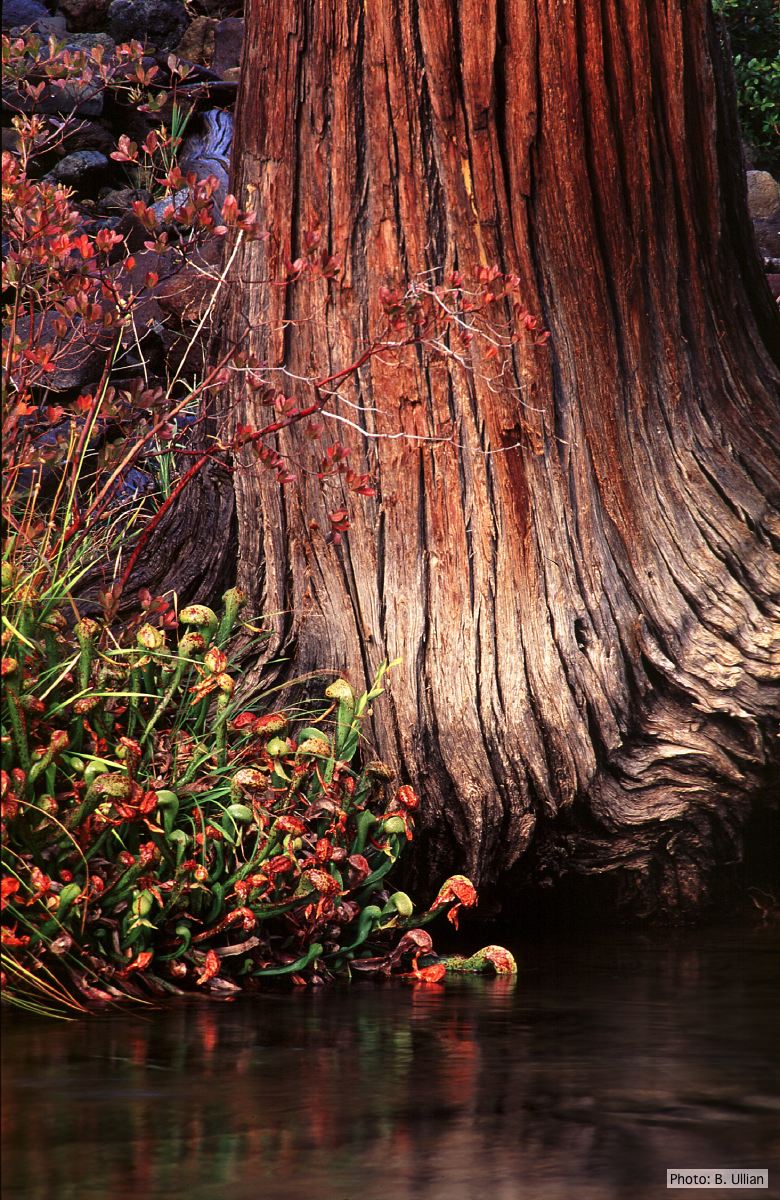 Healthy Chamaecyparis lawsoniana tree |
|
P. pluvialis sporangium 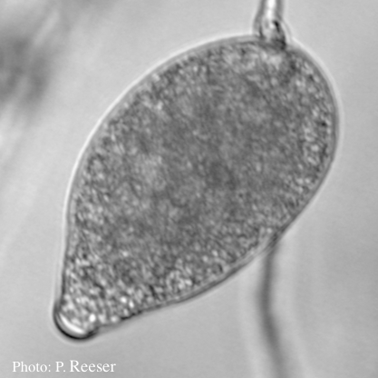 Sporangium showing typical ovoid shape and semi-papillate condition |
Dead Port Orford Cedar trees 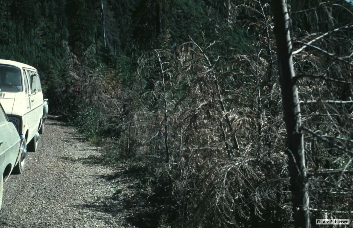 Dead Chamaecyparis lawsoniana trees along road |
P. cambivora sporangia 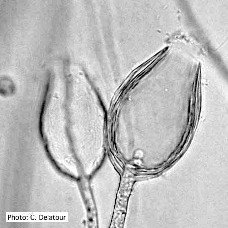 Empty sporangia of P. cambivora showing nested internal proliferation |
|
P. cambivora sporangium 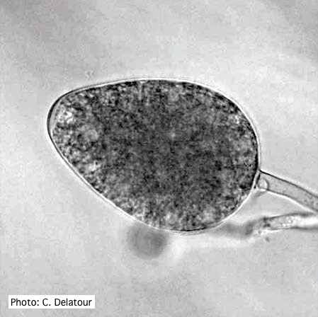 Ovoid non- papillate sporangia |
P. pseudosyringae sporangium 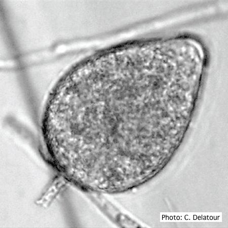 Ovoid, semipapillate sporangia showing medium length pedicel |
P. siskiyouensis canker on Italian alder  Bleeding canker at the base of a tree and a sprinkler emitter (arrow) adjacent to the trunk |