

Colony morphology of P. pinifolia at 20°C on V8 after 3 weeks. From Duran et al. 2008
Photo Gallery
|
P. pinifolia colony morphology on V8 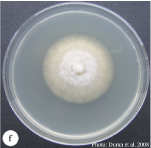 |
P. chlamydospora colony morphology on carrot agar 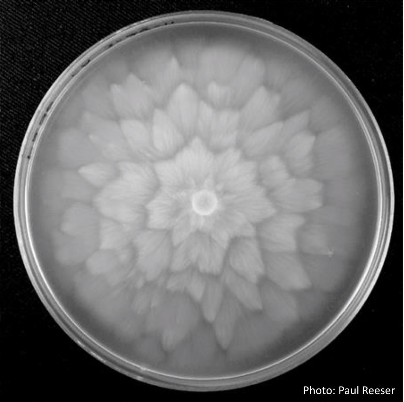 P. chlamydospora colony morphology on carrot agar |
P. agathidicida growth on MEA  Diffuse, non-patterned, colony morphology of ICMP 16471 (the original “Gadgil isolate”) after 10-days incubation at 20°C in the dark |
|
P. cambivora oogonium 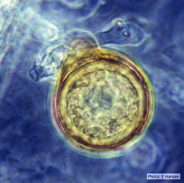 P. cambivora oogonium with antheridium |
Growth of P. arenaria on MEA 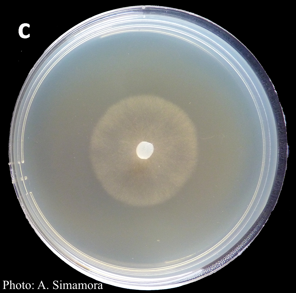 Colony morphology of Phytophthora arenaria after 7 days at 20°C on malt extract agar |
P. cryptogea hyphal swellings 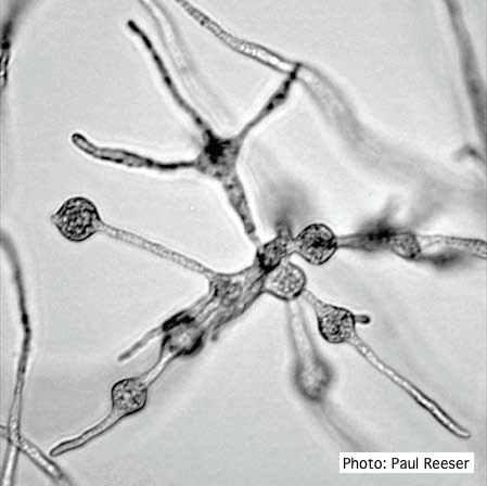 Cluster of small, angular to globose hyphal swellings formed in water |
|
P. cambivora sporangium with sympodial proliferation 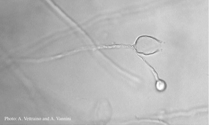 Empty sporagium showing sympodial proliferation |
Dead and healthy Port-Orford cedar seedlings  Port-Orford-cedar seedlings planted to test for Phytophthora lateralis resistance at the Dorena Genetic Resource Center |
P. nicotianae colony morphology on PDA 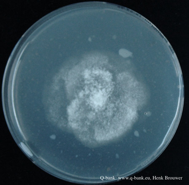 Phytophthora nicotianae CBS 321.49 PDA after 7 days at 24 degrees. Photo from Q-bank: www.q-bank.eu, Henk Brouwer (CBS-KNAW, Utrecht, The Netherlands) |
|
P. agathidicia oogonia 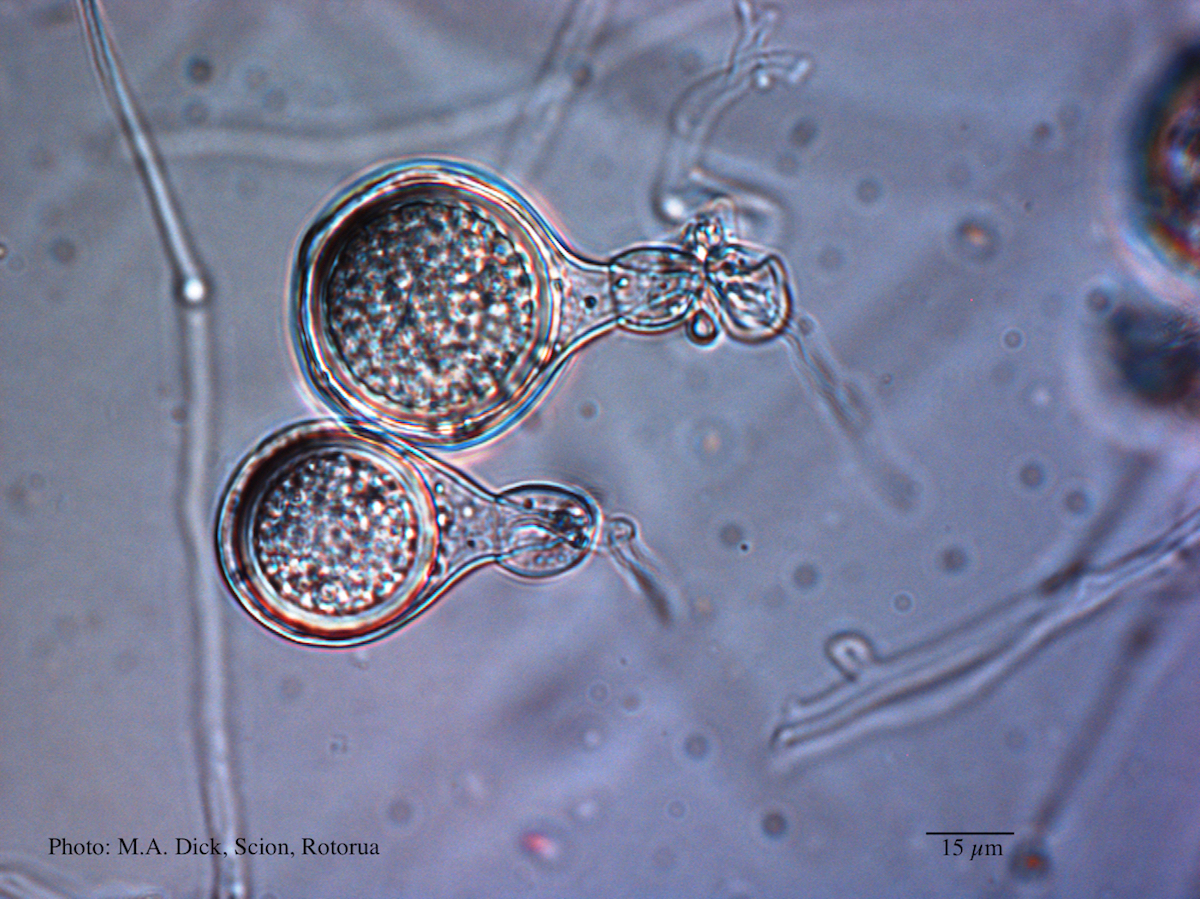 Light micrograph of P. agathidicida oospore (Scale bar equals 15 µm) |
P. megasperma oogonium 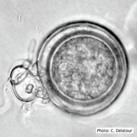 Oogonium with paragynous antheridia applied close to the ogonial stalk. |
P. frigida chlamydospore 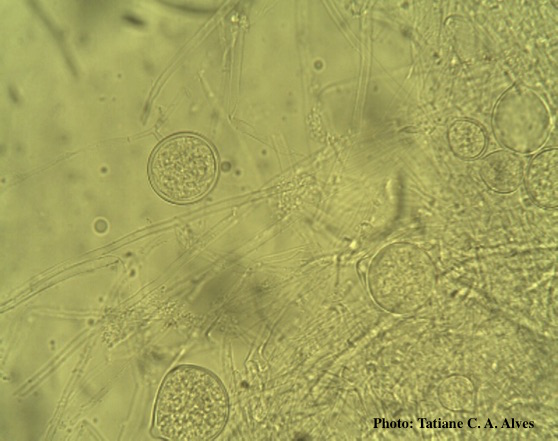 Globose chlamydospores of P. frigida |