P. chlamydospora colony morphology on V8 agar
P. chlamydospora colony morphology on V8 agar
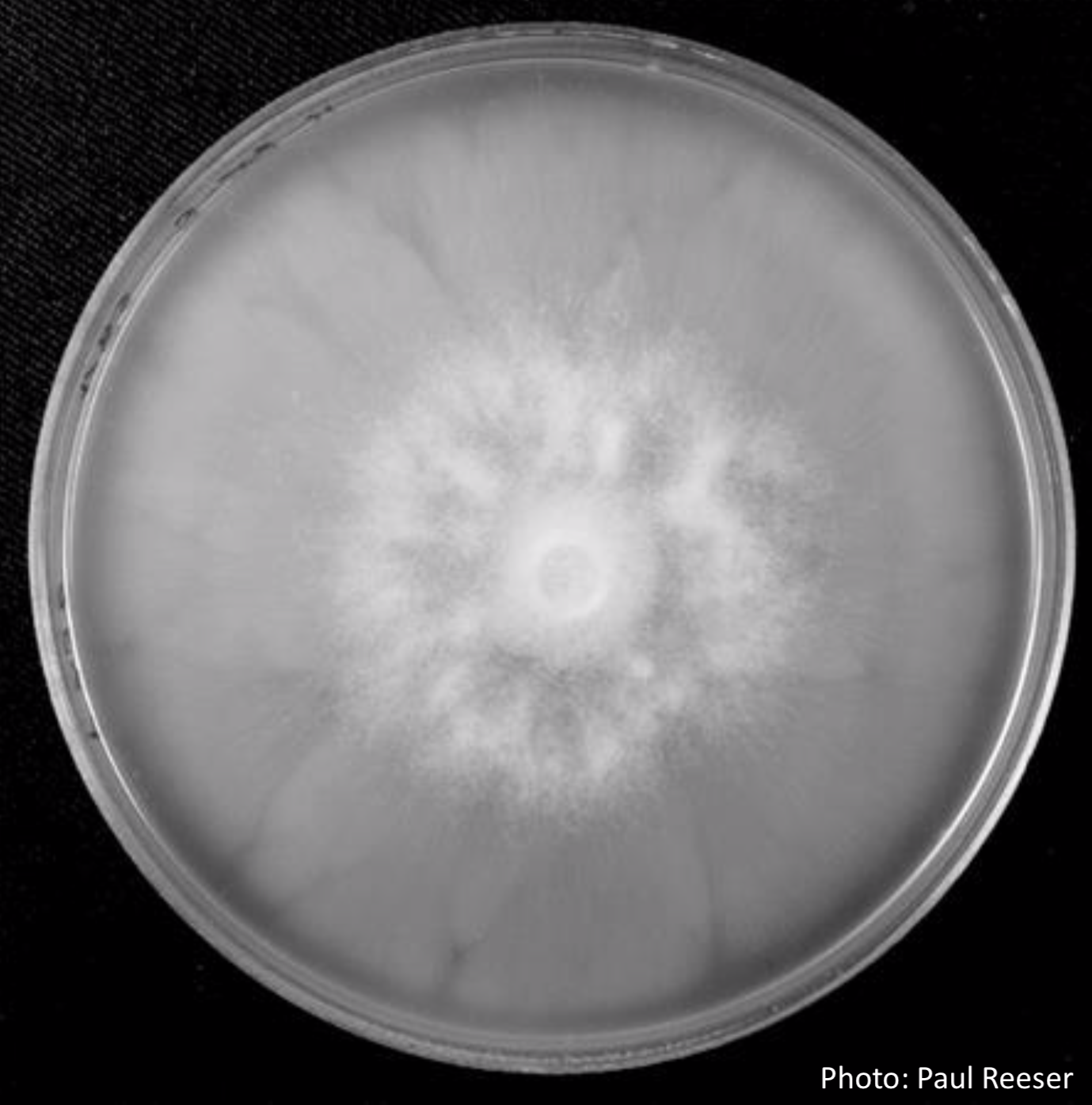
Photographer:
Paul Reeser
Colony Morphology:
All colony morphology
V8 Agar
Scale:
Organismal


P. chlamydospora colony morphology on V8 agar
Phytophthora nicotianae CBS 321.49 CMA after 7 days at 24 degrees. Photo from Q-bank: www.q-bank.eu, Henk Brouwer (CBS-KNAW, Utrecht, The Netherlands)

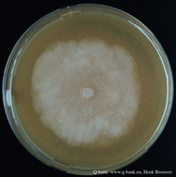
Phytophthora nicotianae CBS 321.49 PDA after 7 days at 24 degrees. Photo from Q-bank: www.q-bank.eu, Henk Brouwer (CBS-KNAW, Utrecht, The Netherlands)
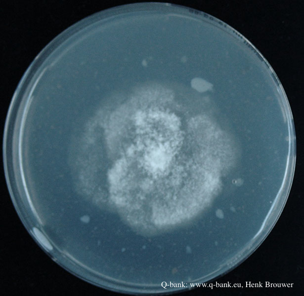
Phytophthora nicotianae CBS 321.49 V8 after 7 days at 24 degrees. Photo from Q-bank: www.q-bank.eu, Henk Brouwer (CBS-KNAW, Utrecht, The Netherlands)
Clusters of sporangia emerge from stomata of an infected radiata pine needle.

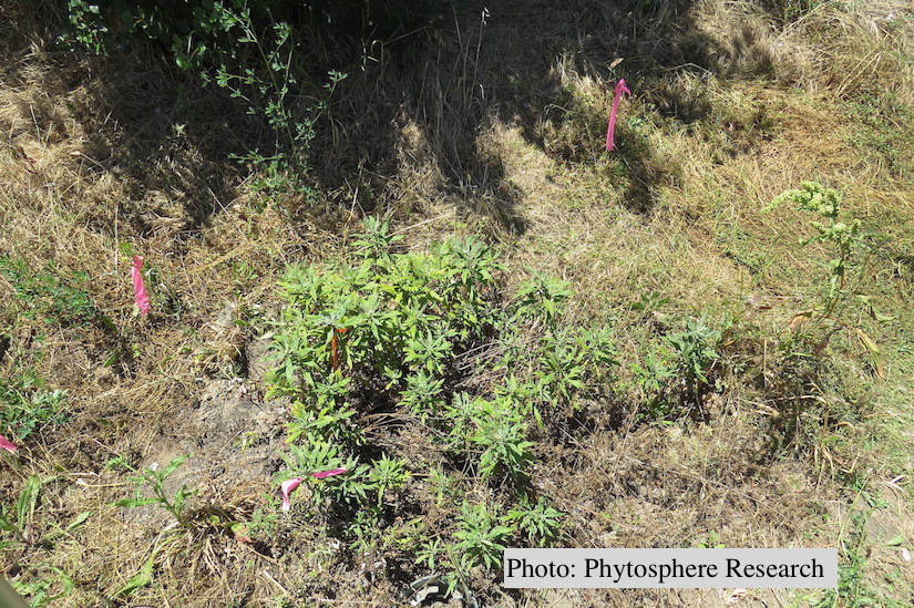
Outplanted California mugwort (Artemisia douglasiana) infected with P. tentaculata, 4.5 years after planting. Plant shows stunting and chlorosis. (P. cryptogea and P. lacustris were also baited from roots/soil of this plant).
Nursery grown California mugwort plant (Artemisia douglasiana) infected with P. tentaculata and exhibiting severe root and crown rot

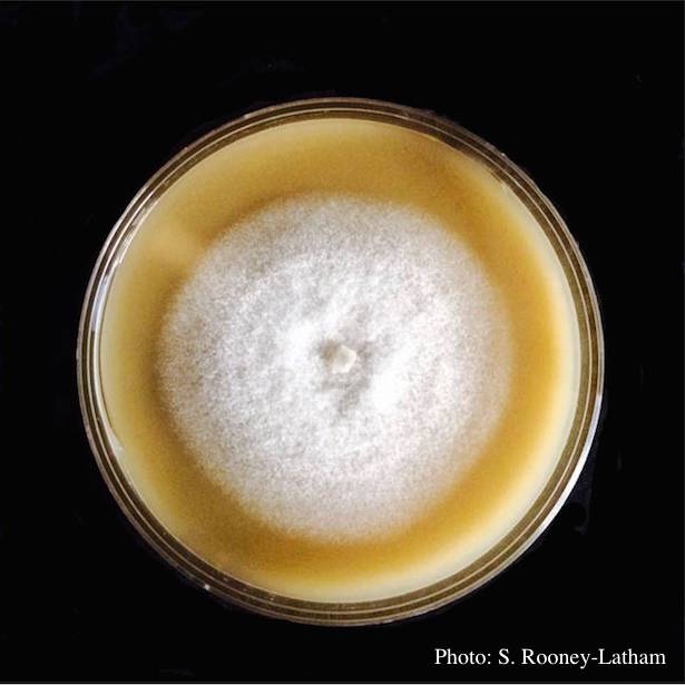
Culture of P. tentaculata on V-8 media
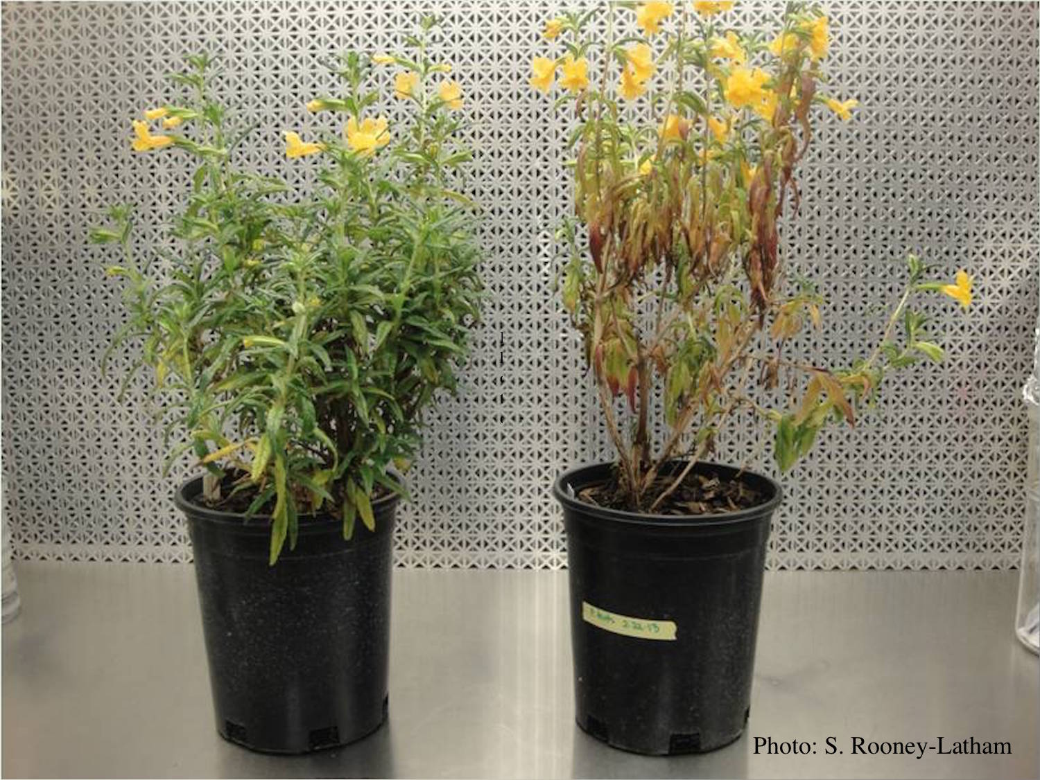
Crown and root rot (left) on sticky monkey flower (Diplacus aurantiacus) compared with a control (right)
Typical red needle cast symptoms along a twig. Lesions begin at the base of the needle which subsequently turns brown and is cast from the twig.
