

Phytophthora chlamydospora chlamydospore in agar. Bar is 20µm.
Photo Gallery
|
P. chlamydospora chlamydospore 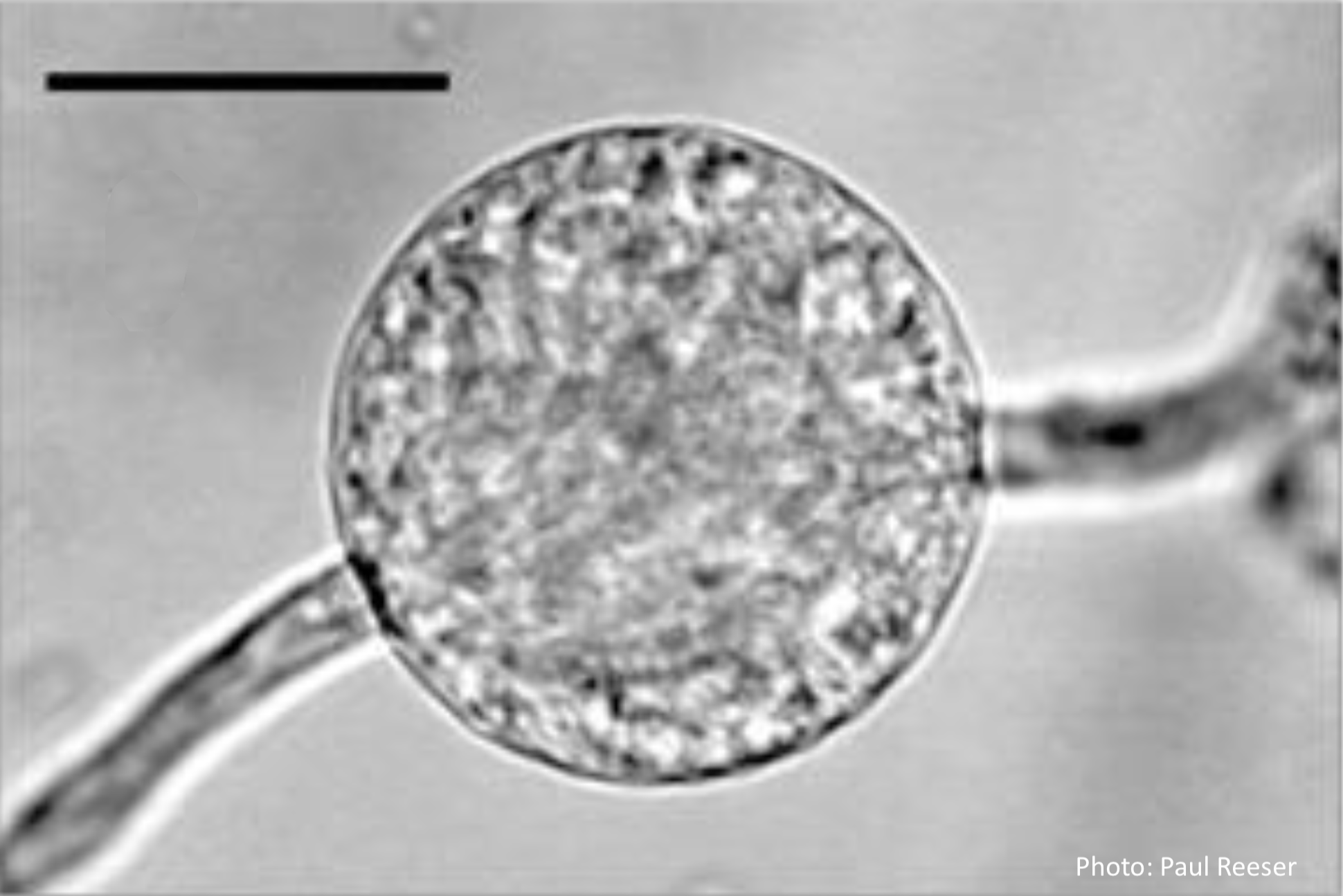 |
P. ramorum bleeding canker 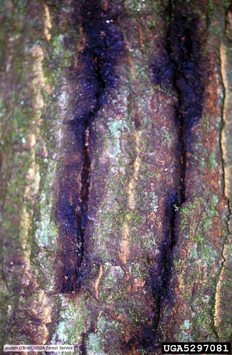 Bark cracks with black ooze in coast live oak, a symptom of P. ramorum canker. |
Stain from Port Orford Cedar root disease  Stain from Chamaecyparis lawsoniana root disease on the Smith River |
|
P. cambivora colony morphology on MA 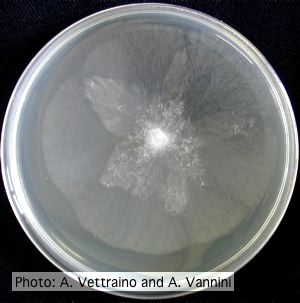 Appressed stellate colony morphology at 14 days at 20°C on MA |
P. siskiyouensis canker on Italian alder  Bleeding canker at the base of a tree and a sprinkler emitter (arrow) adjacent to the trunk |
P. tentaculata oogonia and antheridia 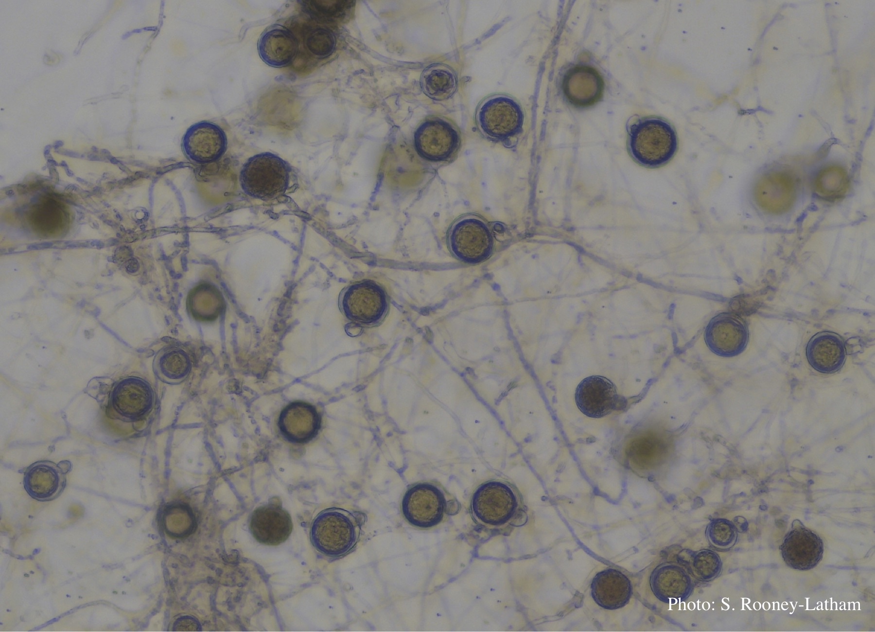 Oospores and oogonia with mostly paragynous but some amphigynous antheridia of P. tentaculata |
|
P. tentaculata microscopic characteristics 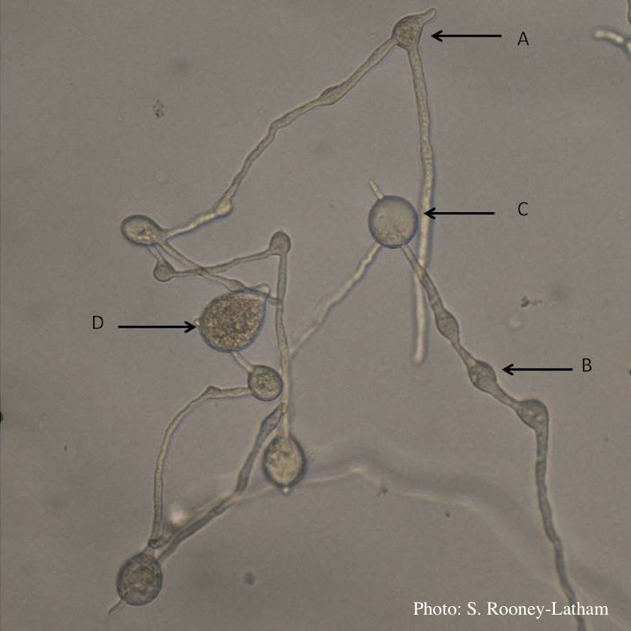 Hyphal swellings occuring at branching points of Mycelium (A), Intercalary hyphal swellings (B), Chlamydospore (C ), Sporangia (D) |
P. cambivora sporangium with sympodial proliferation 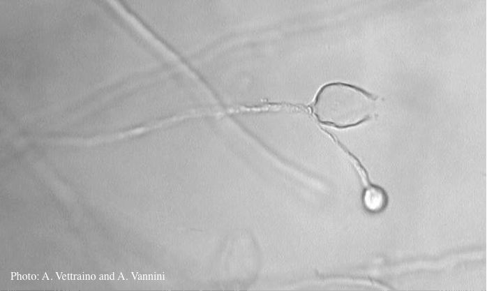 Empty sporagium showing sympodial proliferation |
P. kernoviae sporangium 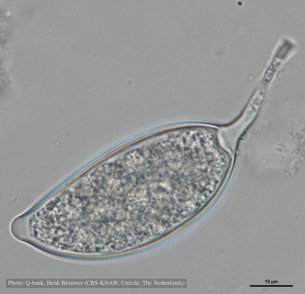 Asymmetrical sporangium, photo from Q-bank, used with permission |
|
P. palmivora chlamydospore 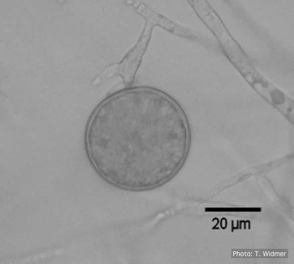 Terminal chlamydospore of P. palmivora |
P. cambivora colony morphology on PDA  Rosaceous colony morphology at 14 days at 20°C on PDA |
P. arenaria disease symptoms on Banksia landscape 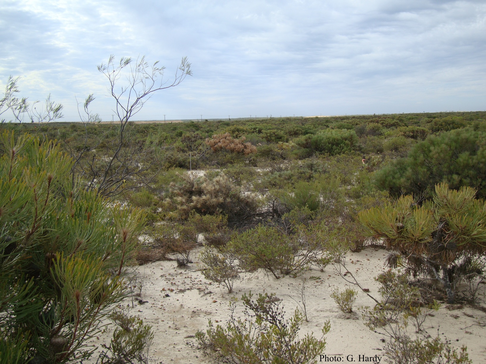 Dead Banksia sp. in a Kwongan heathland on mineral sand near Eneabba, Western Australia recently killed by root and collar rot caused by Phytophthora arenaria |