

Phytophthora chlamydospora sporangia in water, showing internal proliferation. Bar is 20 µm.
Photo Gallery
|
Phytophthora chlamydospora sporangium 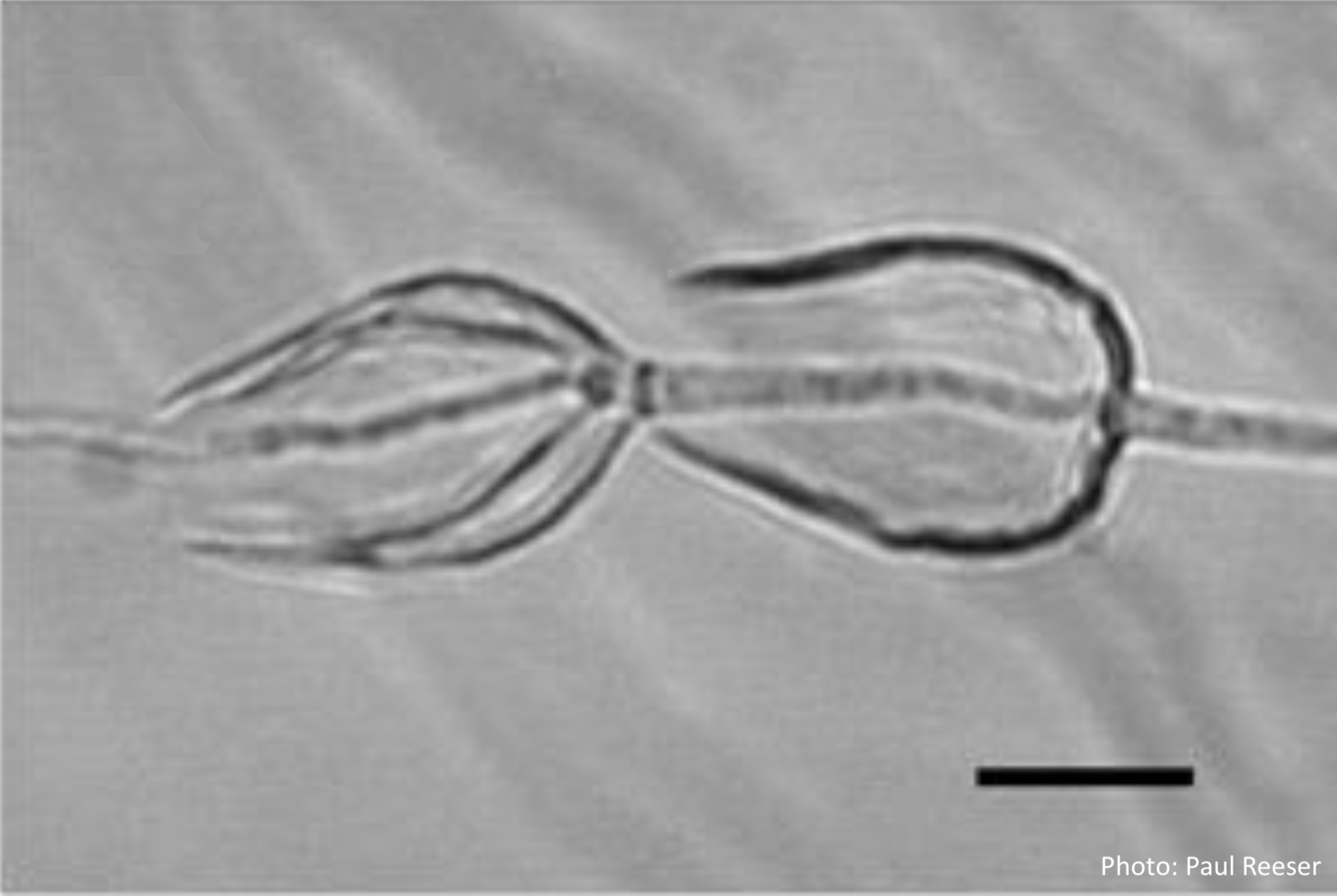 |
P. siskiyouensis colony morphology on PDA 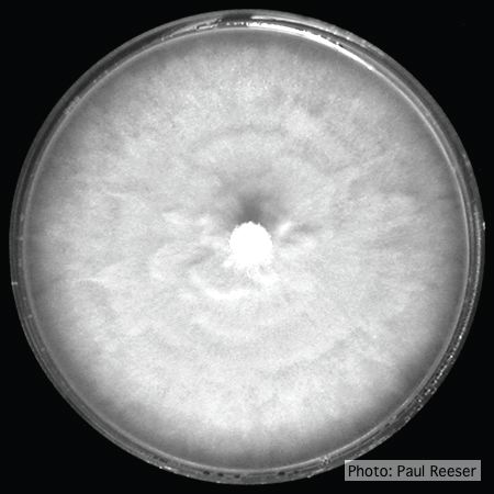 Colony morphology on PDA at 14 days |
P. austrocedrae hyphal swellings in liquid media drawing  Morphology of hyphae of Phytophthora austrocedrae, from Greslebin et al. 2007 |
|
P. cambivora oogonium 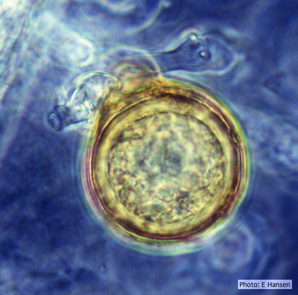 P. cambivora oogonium with antheridium |
Phytophthora cactorum canker on beech tree  Canker on Fagus sp. caused by Phytophthora cactorum. |
P. pinifolia on Pinus radiata  Pinus radiata dead needles caused by DFP with healthy new growth from DFP |
|
P. tentaculata oogonia and antheridia 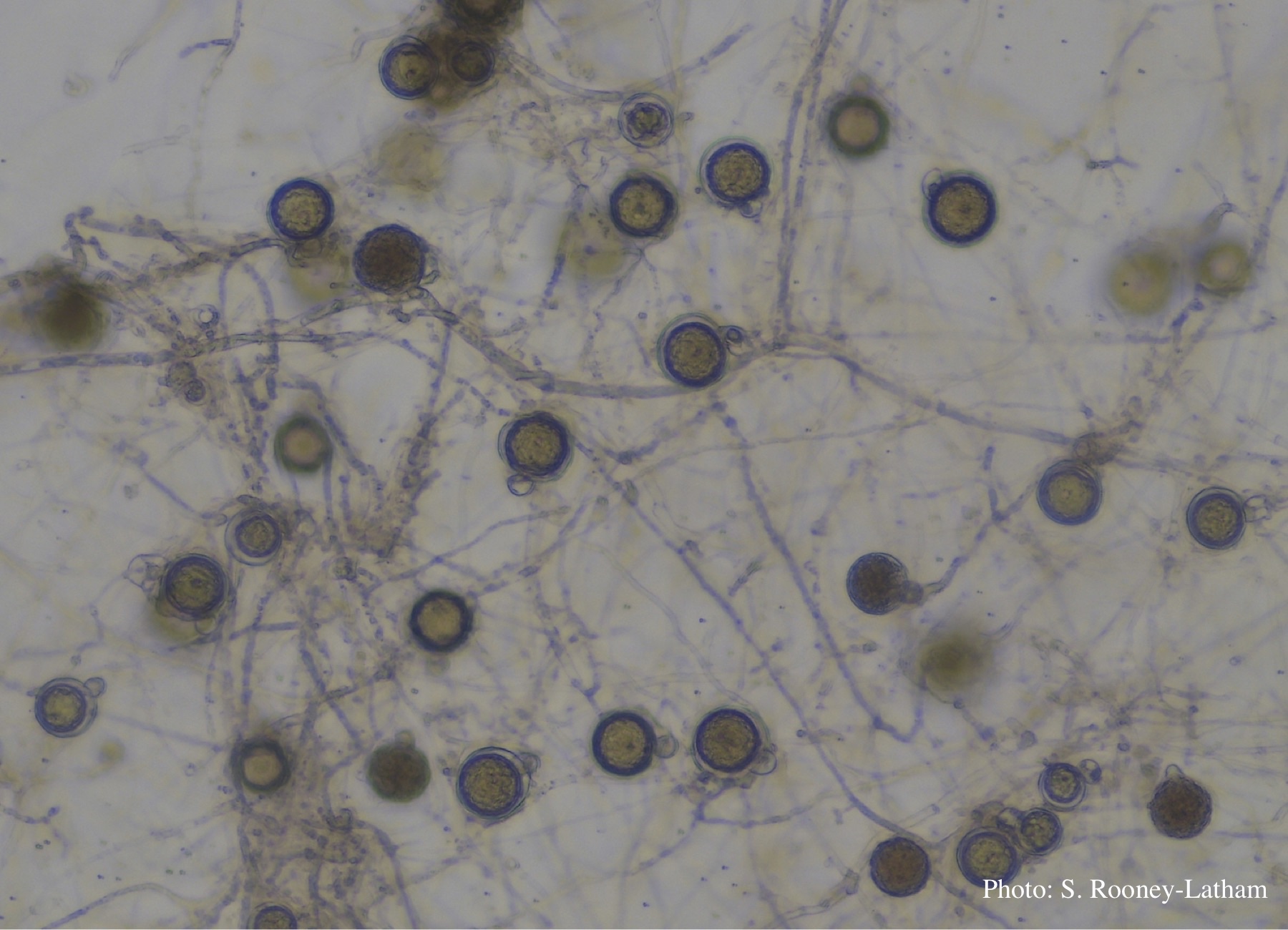 Oospores and oogonia with mostly paragynous but some amphigynous antheridia of P. tentaculata |
P. cactorum sporangia 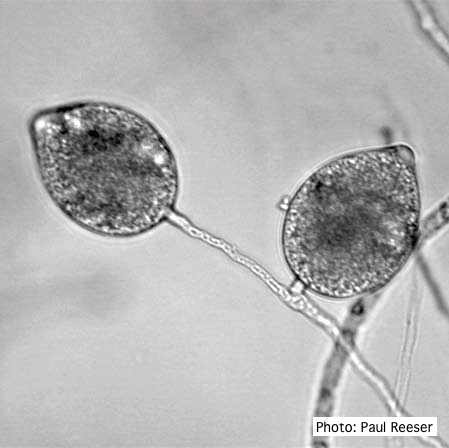 Broadly ovoid, papillate sporangia in water. |
P. alni in alder forest, Germany, with T. Jung 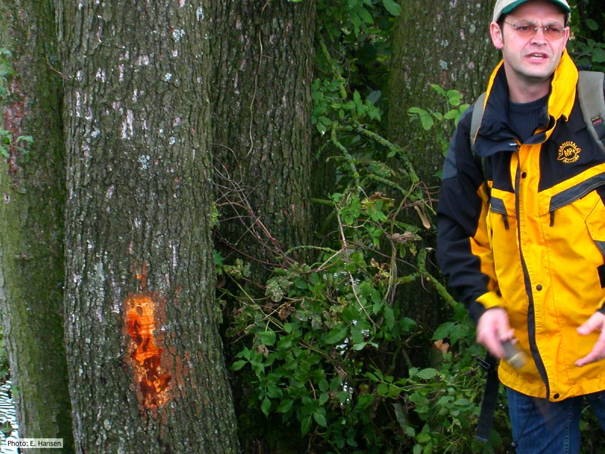 P. alni in alder forest, Germany, with T. Jung |
|
P. cambivora colony morphology on PDA 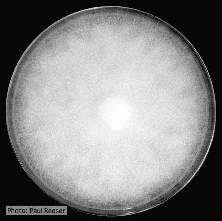 Colony morphology on PDA at 14 days |
P. ramorum oogonium 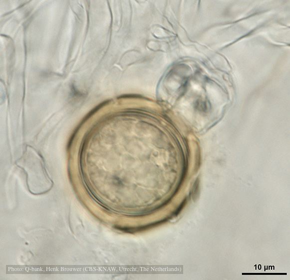 Oogonium with thick oogonial wall, photo from Q-bank, used with permission |
P. austrocedrae - Mal del ciprés, stages of decline 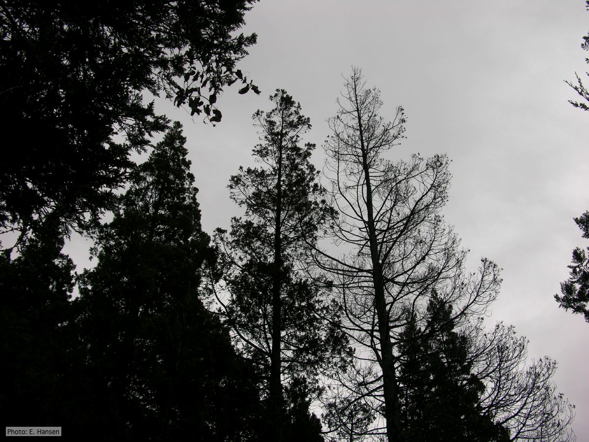 Mal del ciprés, stages of decline |