

Sporangia (sporangiospores), chlamydospores, and hyphae stained with Cotton Blue
Photo Gallery
|
P. palmivora sporangia, chlamydospores, hyphae 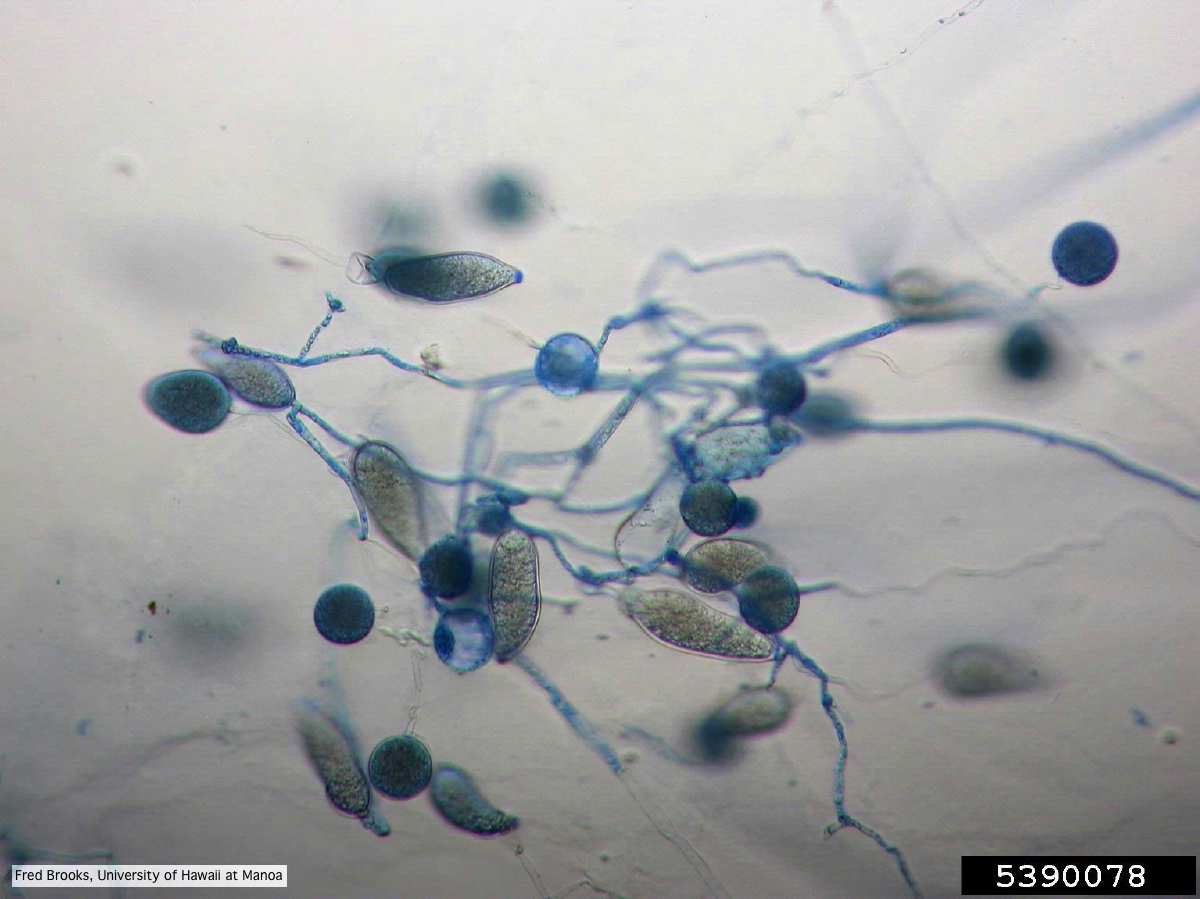 |
P. lateralis on Port Orford cedar  Dying Chaemacyparis lawsoniana trees in Lopérec, France. |
P. pluvialis hyphal swellings 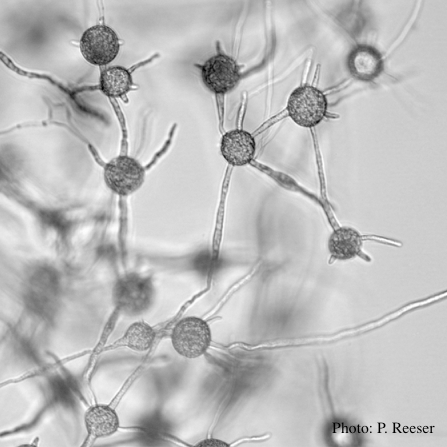 P. pluvialis hyphal swellings in water |
|
P. siskiyouensis canker on Italian alder  Bleeding canker at the base of a tree and a sprinkler emitter (arrow) adjacent to the trunk |
P. austrocedrae colony morphology on Tomato juice agar with B sitosterol 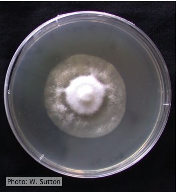 Colony morphology of P. austrocedrae at 16 ºC after 4 weeks on Tomato juice agar with B sitosterol |
P. frigida oogonium  Oogonium and oospore with amphigynous antheridium |
|
P. ramorum sporangium 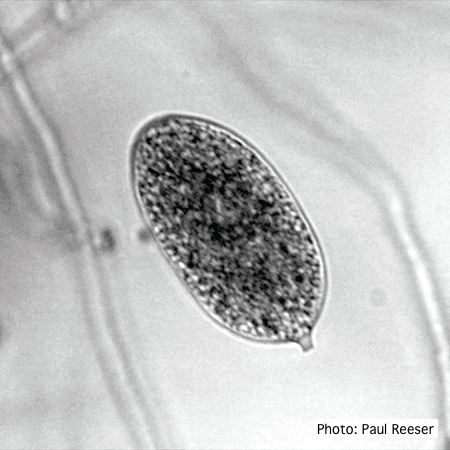 P. ramorum sporangium |
Growth morphology on V8 of P. lateralis 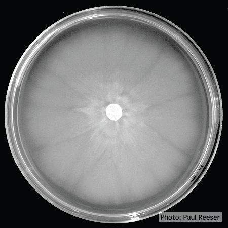 Colony morphology on V8 at 14 days |
P. tentaculata chlamydospore  P. tentaculata chlamydospore with short hyphal projection |
|
Comparative gametangial morphology of Phytophthora Clade 5 species  Comparative gametangial morphology of Phytophthora Clade 5 species, with SEM (top) and light microscopy (bottom). P. heveae has smooth walled oogonia with funnel-shaped, amphigynous antheridia. P. agathidicida has mildly stipulate oogonia with globose amphigynous antheridia. P.cocois has mildly bullate oogonia with reflexed amphigynous antheridia. P. castaneae has coarsely bullate oogonium with rugose protuberances and narrow amphigynous antheridia (Weir et al. 2015). |
Healthy Port Orford Cedar tree 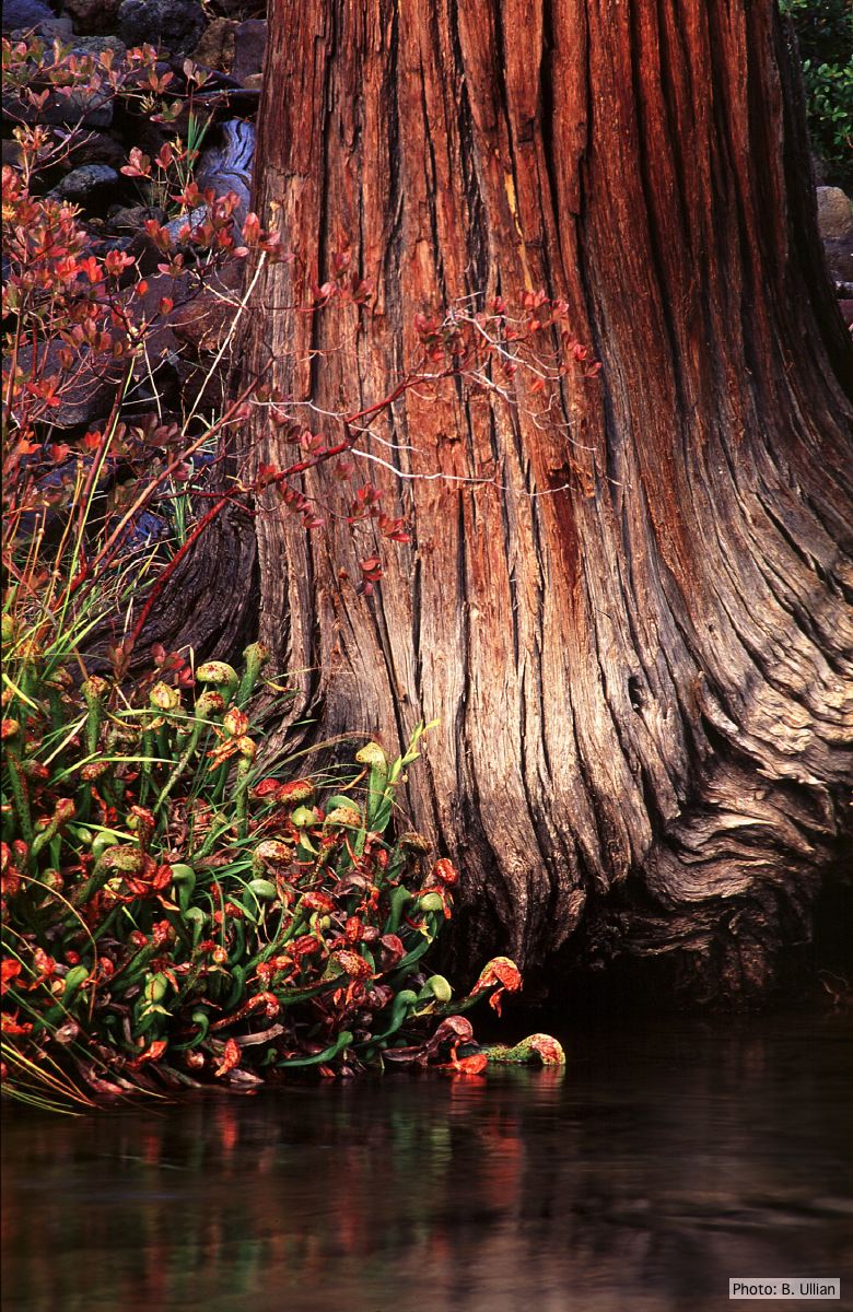 Healthy Chamaecyparis lawsoniana tree |
P. cactorum bleeding canker  Bleeding canker on red oak (Quercus rubra) |