

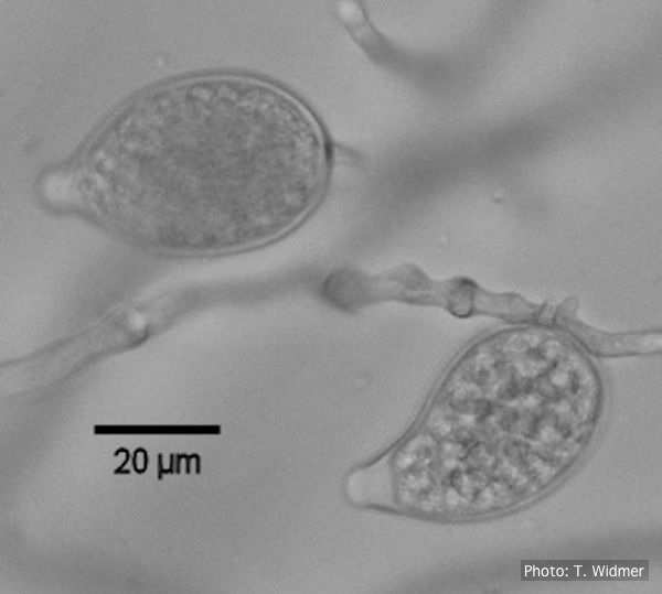
P. nicotianae overview of sporangia 40x. Photo from Q-bank: www.q-bank.eu, Henk Brouwer (CBS-KNAW, Utrecht, The Netherlands)
Photo Gallery
|
P. nicotianae sporangia  |
P. agathidicida oospores  Oospores of P. agathidicida in the roots of kauri seedlings inoculated with P. agathidicida. The root has been cleared with potassium hydroxide and bleached with peroxide, before being stained with Trypan Blue |
P. frigida chlamydospore 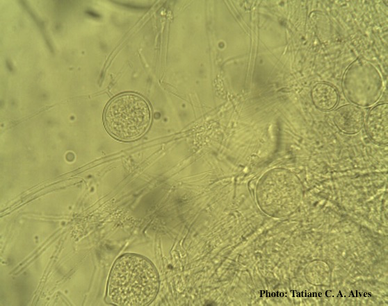 Globose chlamydospores of P. frigida |
|
Basal canker on Port-Orford cedar 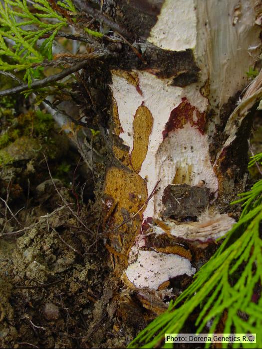 Basal canker on Chamaecyparis lawsoniana |
P. siskiyouensis bleeding canker  Bole lesions in the tissues under the bark of a bleeding canker: discoloration in the secondary phloem tissue |
P. agathidicida lesion on kauri tree 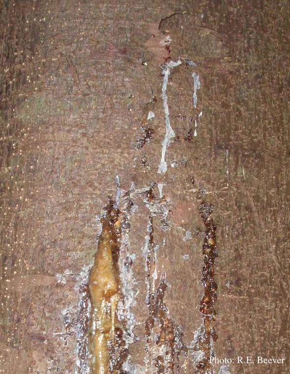 Gum oozing out of longitudinal lesion |
|
P. cinnamomi colony morphology on PDA  P. cinnamomi colony growth on PDA at 14 days |
Dead Port Orford Cedar trees 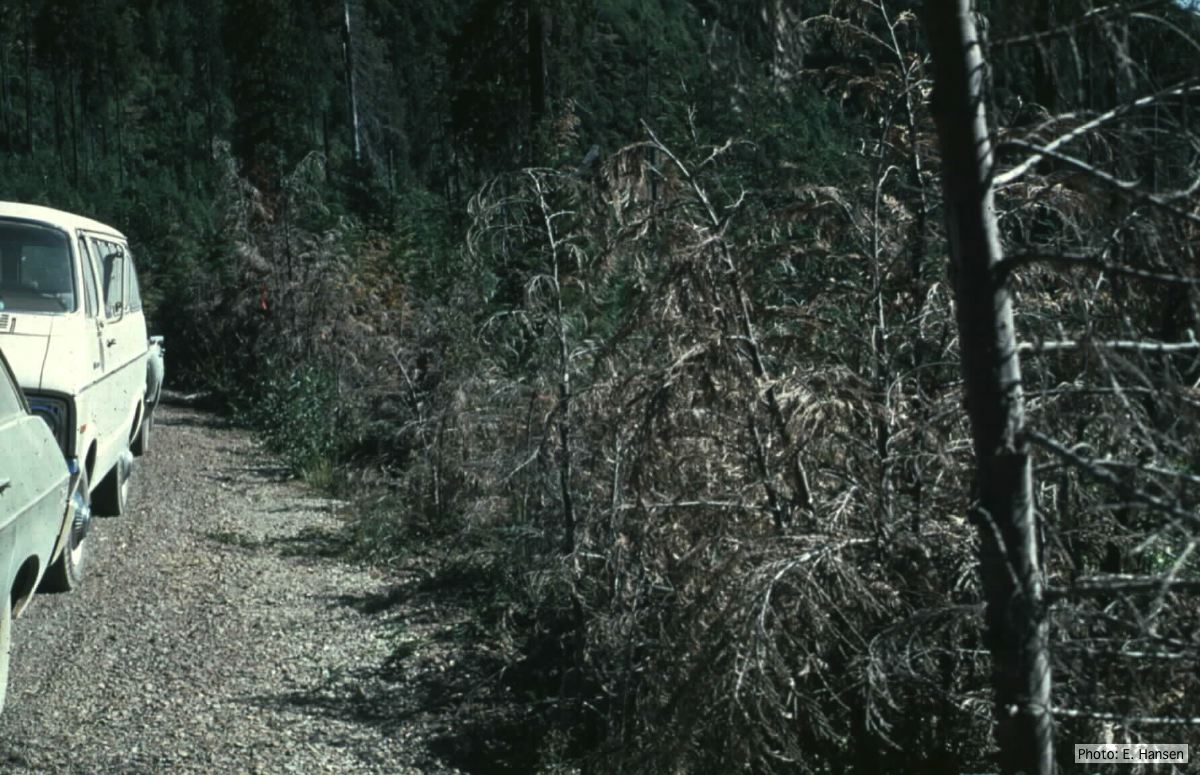 Dead Chamaecyparis lawsoniana trees along road |
P. cambivora colony morphology on PDA 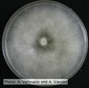 Uniform fluffy colony morphology at 14 days at 20°C on PDA |
|
P. lateralis on Port Orford cedar 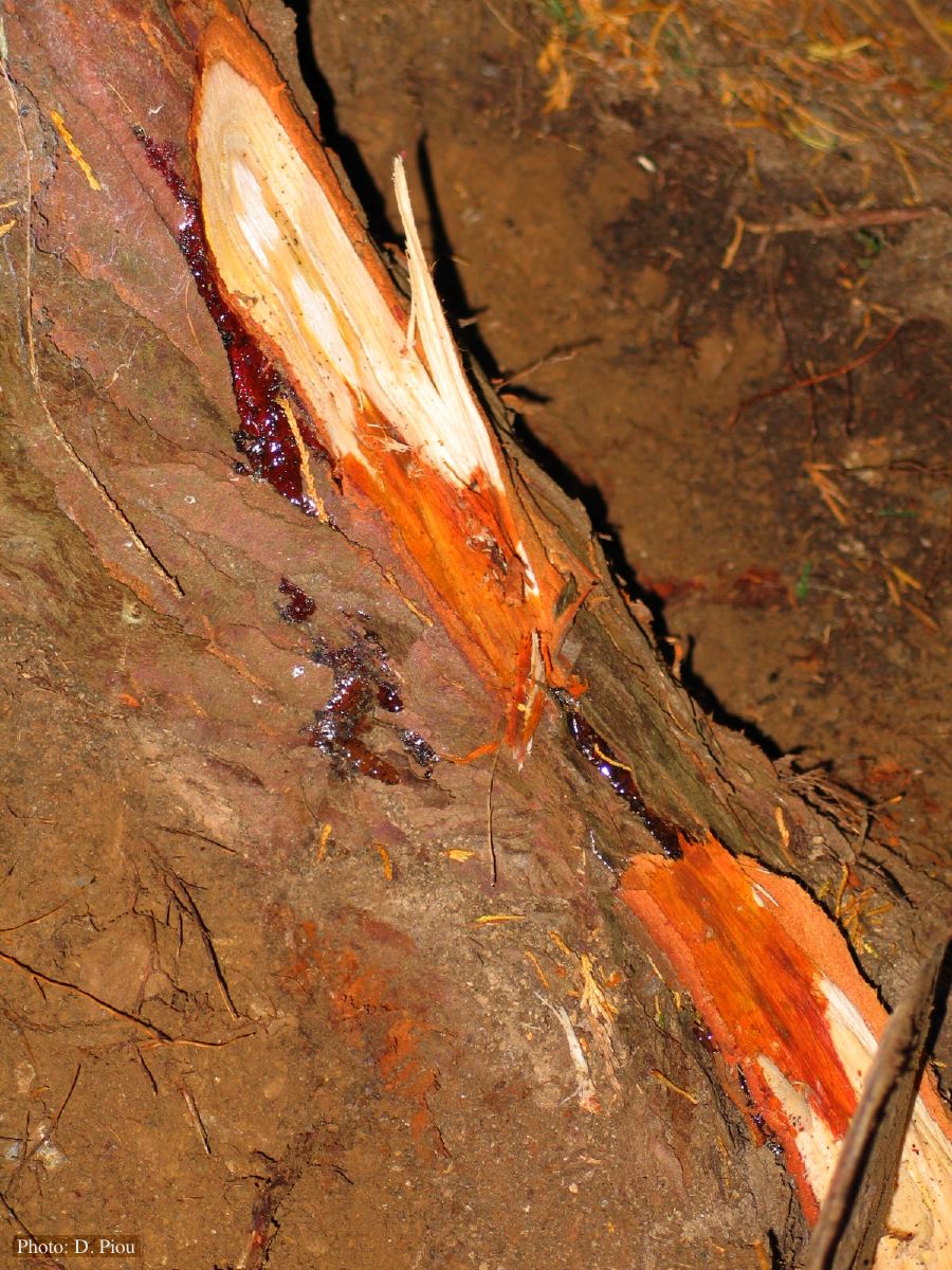 Small root lesions on Chaemacyparis lawsoniana |
P. nicotianae colony morphology on V8 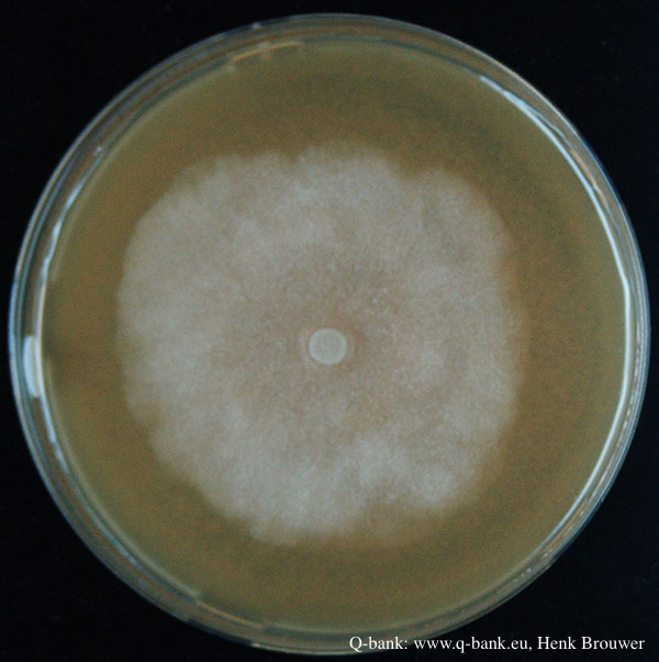 Phytophthora nicotianae CBS 321.49 V8 after 7 days at 24 degrees. Photo from Q-bank: www.q-bank.eu, Henk Brouwer (CBS-KNAW, Utrecht, The Netherlands) |
P. palmivora sporangia
 P. palmivora caducous papillate sporangia
|