

Laterally intercalary chlamydospore of Phytophthora lateralis
Photo Gallery
|
Chlamydospore of P. lateralis 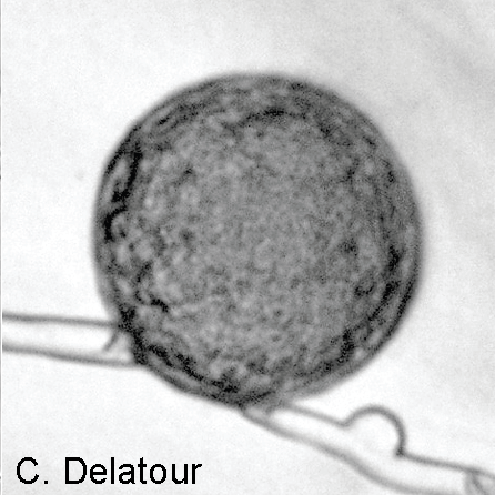 |
P. cambivora disease symptoms 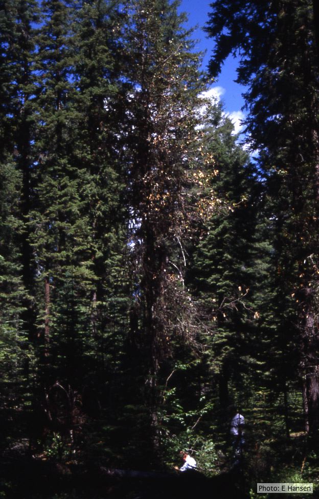 Dead and dying chinquapin infected with P. cambivora |
P. palmivora colony morphology on V8 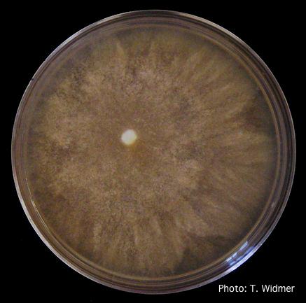 Growth of P. palmivora on V8 agar |
|
P. cactorum bleeding canker 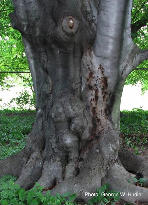 Bleeding canker on European beech (Fagus sylvatica) |
P. cactorum colony morphology on PDA 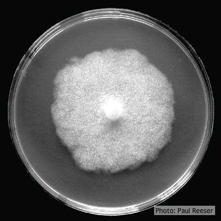 Colony morphology on PDA at 14 days |
P. nemorosa oogonium 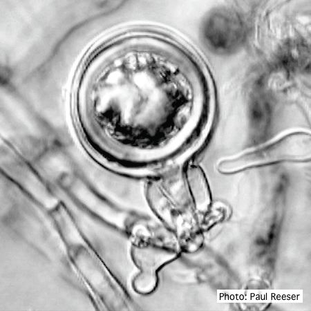 Oogonium with amphigynous antheridium |
|
P. pinifolia on Pinus radiata 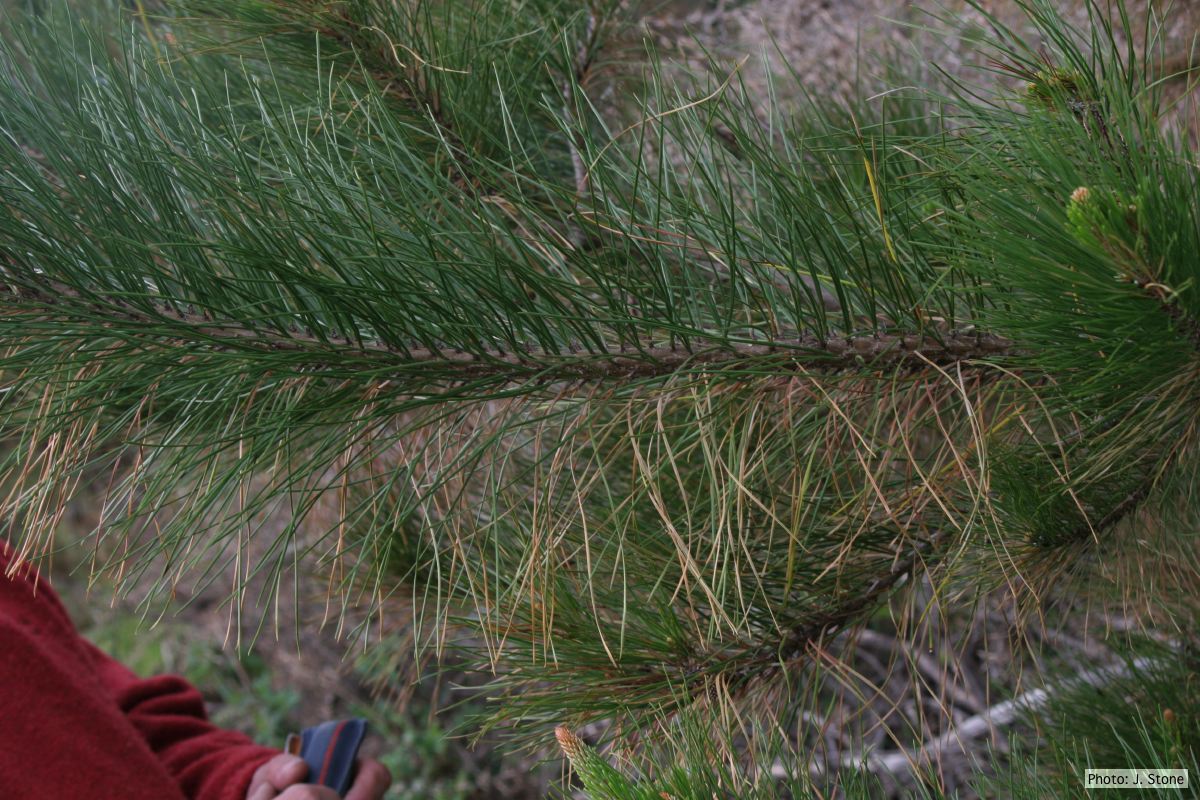 Dead needles on lower side of P. radiata branch. |
P. cambivora tar spots 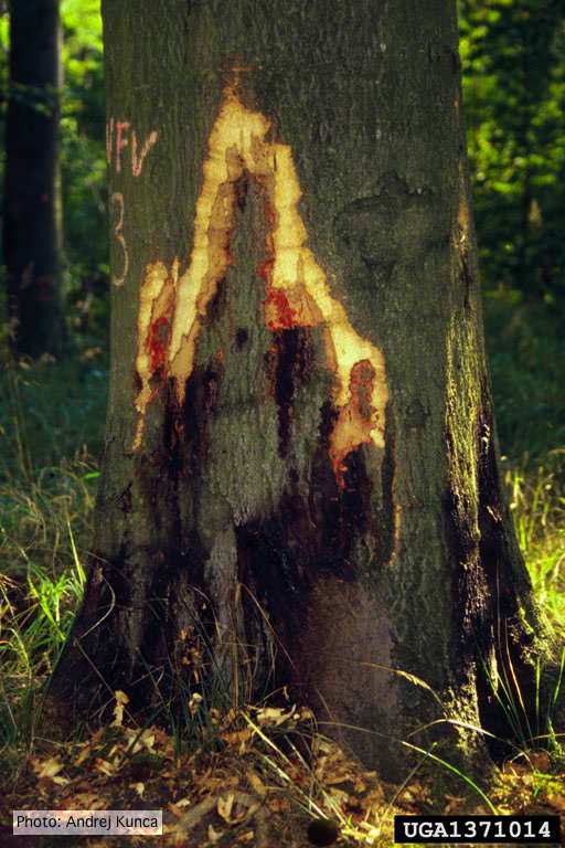 Tar spots on European beech (Fagus sylvatica) with bark removed. Lesse, Germany |
P. katsurae oogonium 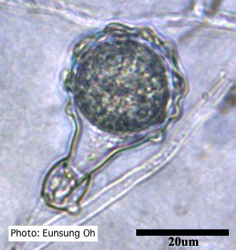 Warty protuberances on oogonium |
|
P. cryptogea colony morpholgy on PDA 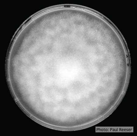 Colony morphology on PDA at 14 days |
Dying Port Orford Cedar trees 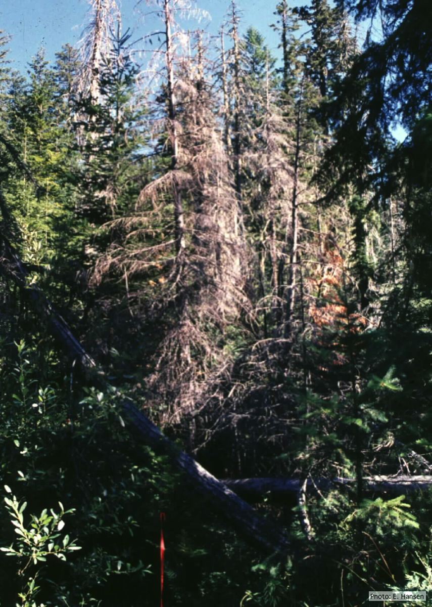 Dead Chamaecyparis lawsoniana trees |
P. agathidicida growth on MEA  Diffuse, non-patterned, colony morphology of ICMP 16471 (the original “Gadgil isolate”) after 10-days incubation at 20°C in the dark |