

Uniform fluffy colony morphology at 14 days at 20°C on PDA
Photo Gallery
|
P. cambivora colony morphology on PDA 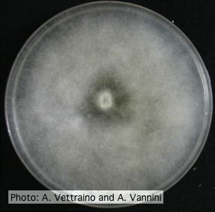 |
P. agathidicia growth on PDA 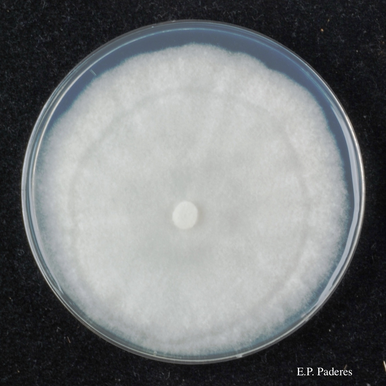 Colony morphology of ex-holotype ICMP 17027 after 10-days incubation at 20°C in the dark |
P. pinifolia on Pinus radiata 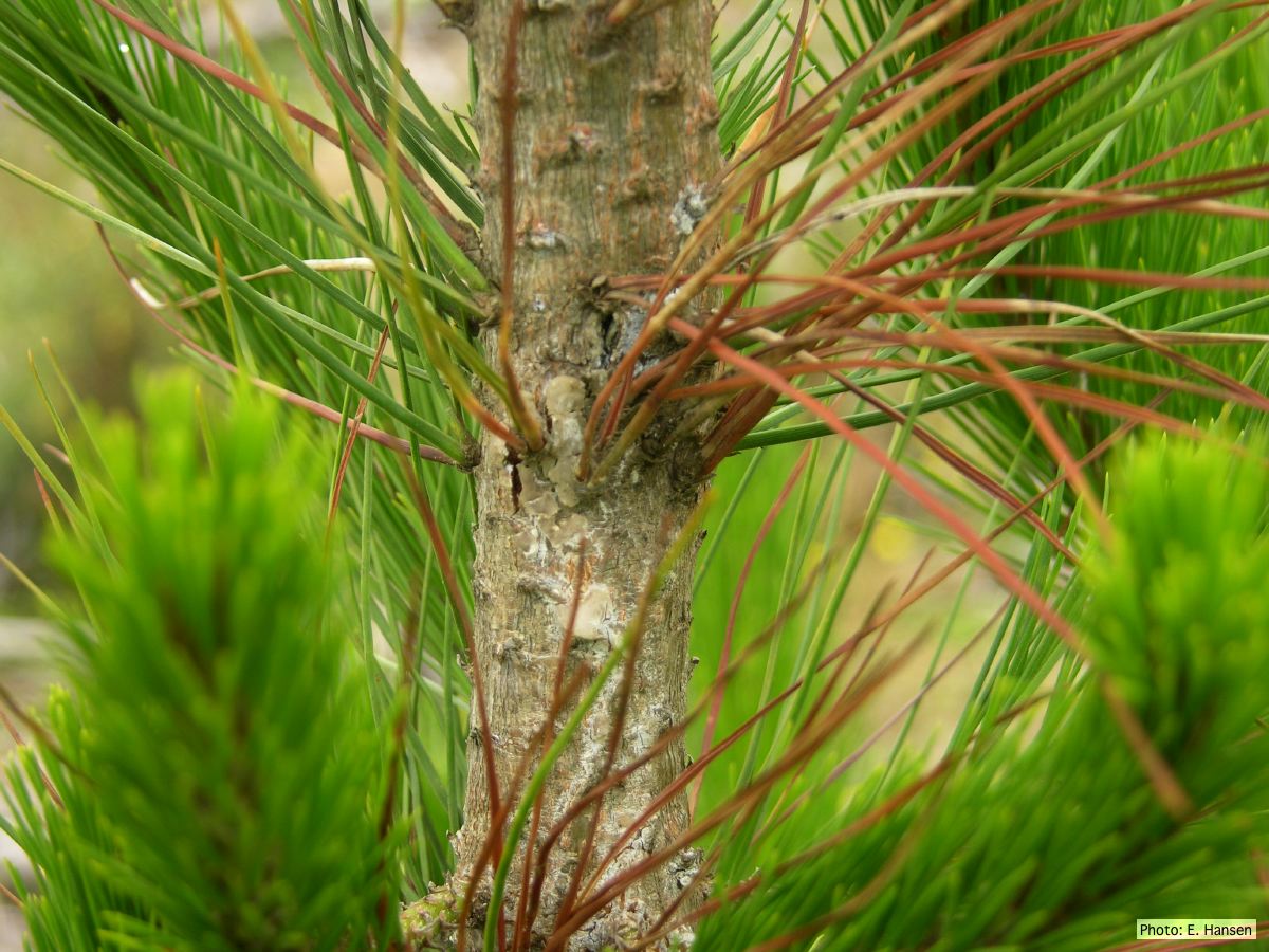 Pinus radiata, note Stem canker associated with necrotic needles. |
|
Basal canker on Port-Orford cedar 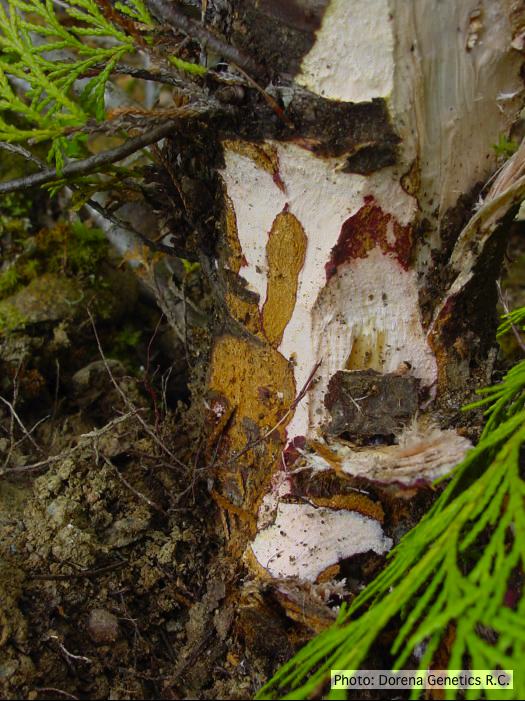 Basal canker on Chamaecyparis lawsoniana |
P. alni canker on gray alder 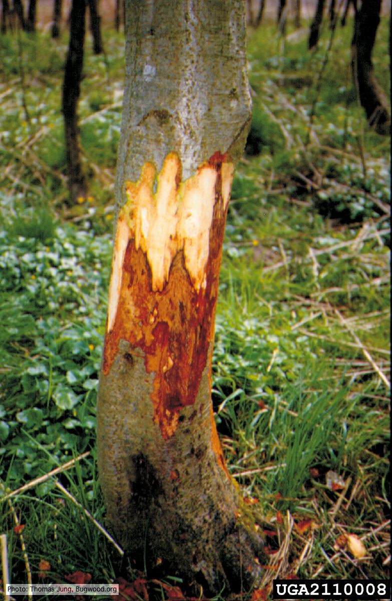 Grey alder (A. incana) with collar rot caused by P. alni |
Chlamydospore of P. lateralis 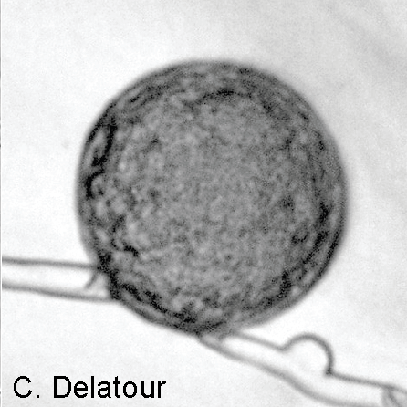 Laterally intercalary chlamydospore of Phytophthora lateralis |
|
P. austrocedrae colony morphology on Tomato juice agar with B sitosterol 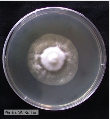 Colony morphology of P. austrocedrae at 16 ºC after 4 weeks on Tomato juice agar with B sitosterol |
P. katsurae oogonium 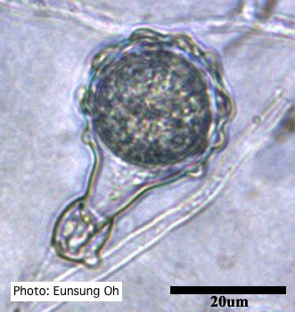 Warty protuberances on oogonium |
Mal del ciprés, dead and dying trees along river 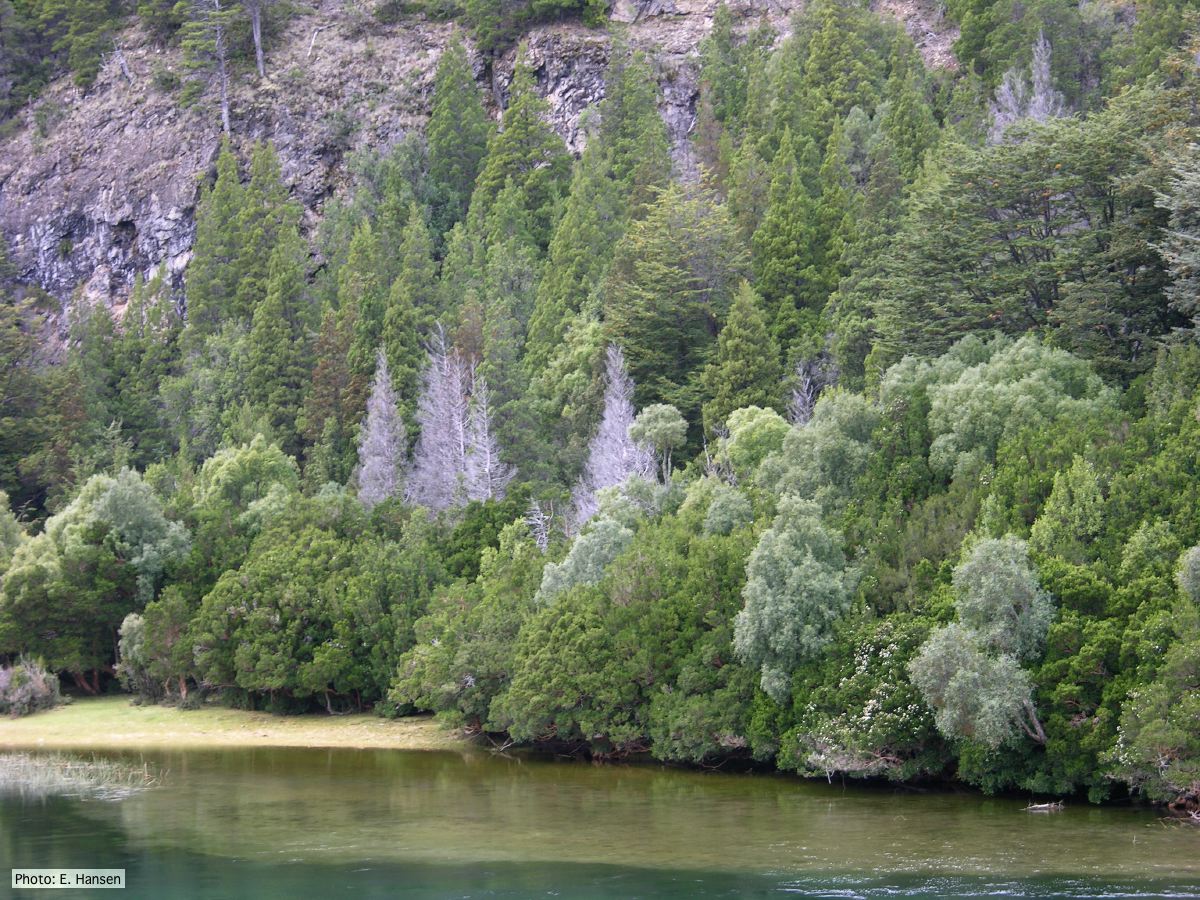 Mal del ciprés, dead and dying trees along river |
|
P. pinifolia on Pinus radiata  Pinus radiata dead needles caused by DFP with healthy new growth from DFP |
P. agathidicia sporangia 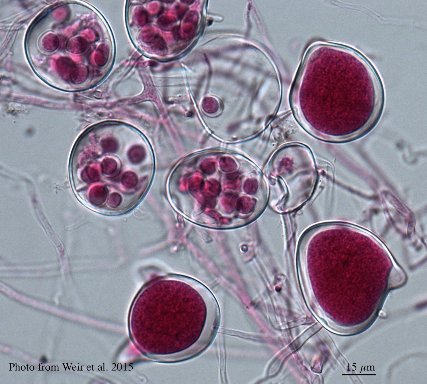 Differentiation of the cytoplasm within papillate sporangia into acid fuchsin stained zoospores |
P. chlamydospora chlamydospore  Phytophthora chlamydospora chlamydospore in agar. Bar is 20µm. |