

Oospores of P. agathidicida in the roots of kauri seedlings inoculated with P. agathidicida. The root has been cleared with potassium hydroxide and bleached with peroxide, before being stained with Trypan Blue
Photo Gallery
|
P. agathidicida oospores  |
P. pinifolia colony morphology on V8 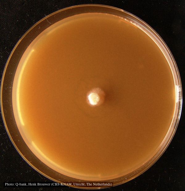 Colony pattern after 7 days on V8 at 24C, photo from Q-bank, used with permission |
Black pod disease of cacao 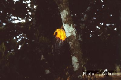 Symptom of black pod disease of cacao (T. cacao) caused by P. palmivora |
|
P. ramorum colony morphology on CMA PARP 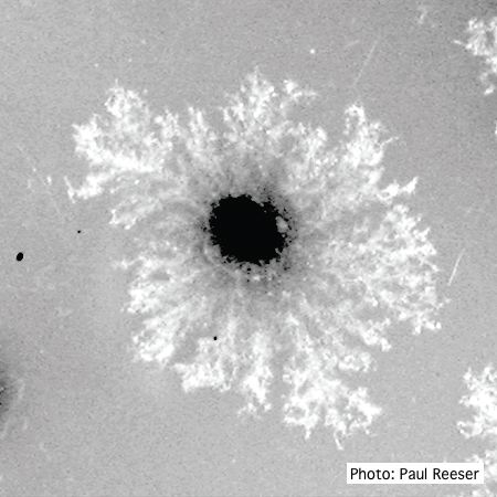 P. ramorum colony morphology on CMA PARP |
P. cambivora oogonium  Bullate oogonium and and two-celled amphigynous antheridium |
Stain from Port Orford Cedar root disease  Stain from Chamaecyparis lawsoniana root disease on the Smith River |
|
P. katsurae sporangia 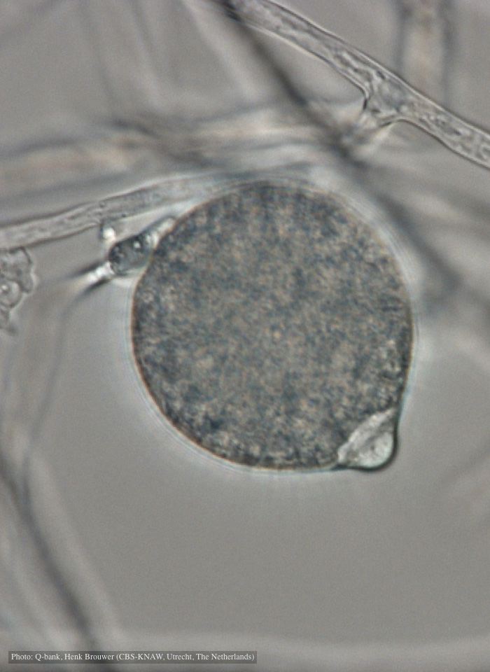 Papillate, non-caducous sporangium with differentiated content; photo used with permission from Q-bank |
P. alni basal canker on European Alder 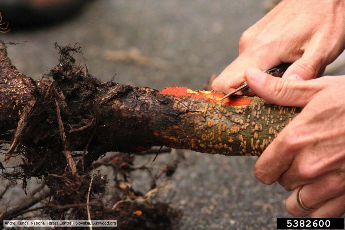 P. alni basal canker on European Alder (Alnus glutinosa) |
P. ramorum oogonium 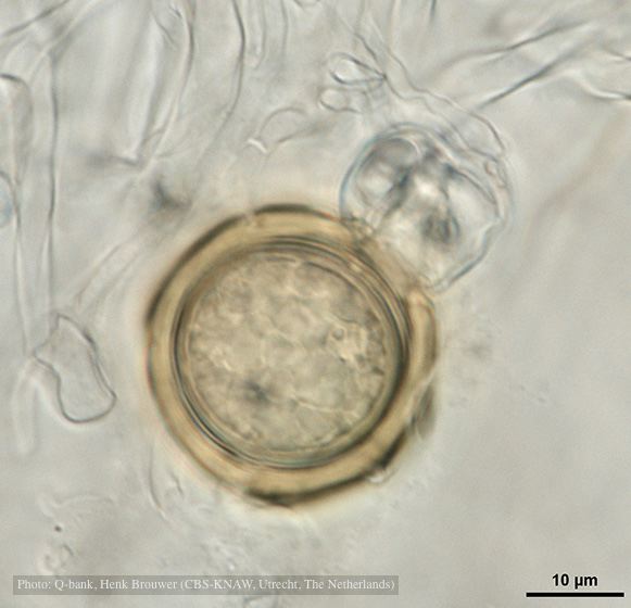 Oogonium with thick oogonial wall, photo from Q-bank, used with permission |
|
P. pinifolia hyphal growth 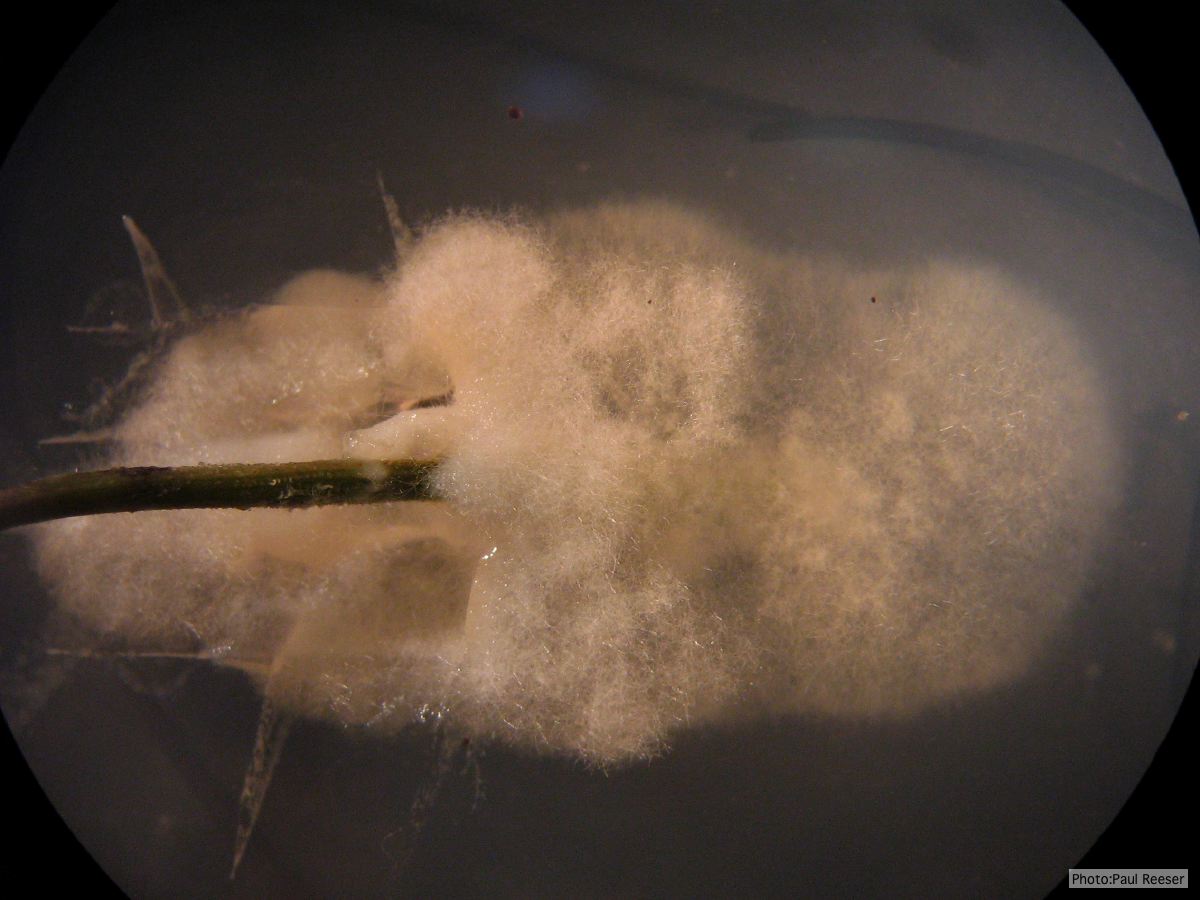 P. pinifolia pathogen growing from infected needle on selective agar |
P. cryptogea sporangium  Ovoid non-papillate sporangia in water. |
P. kernoviae oogonia 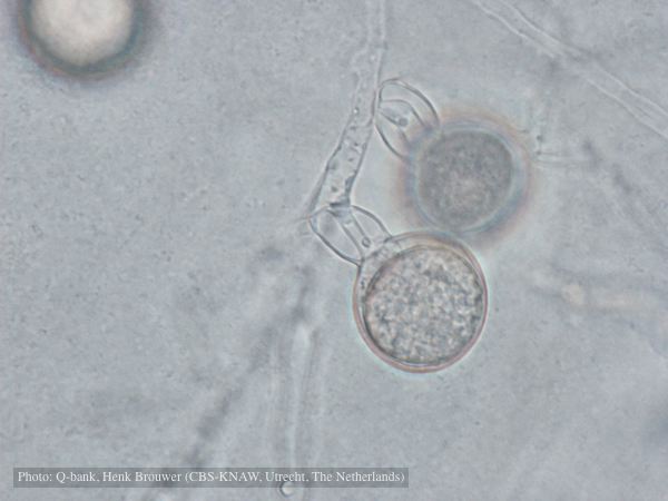 Oogonium with amphigynous antheridia, photo from Q-bank, used with permission |