

Colony morphology on PDA at 14 days
Photo Gallery
|
P. nemorosa colony morphology on PDA  |
P. pinifolia colony morphology on PDA 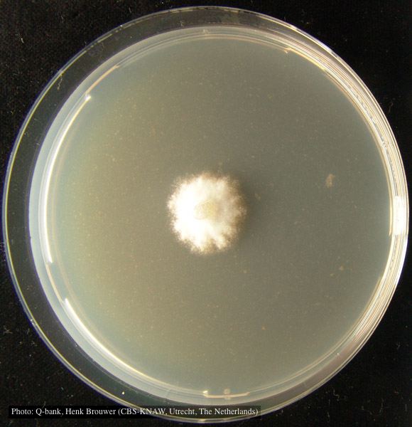 Colony pattern after 7 days on PDA at 24C, photo from Q-bank, used with permission. |
P. pseudotsugae colony morphology on V8 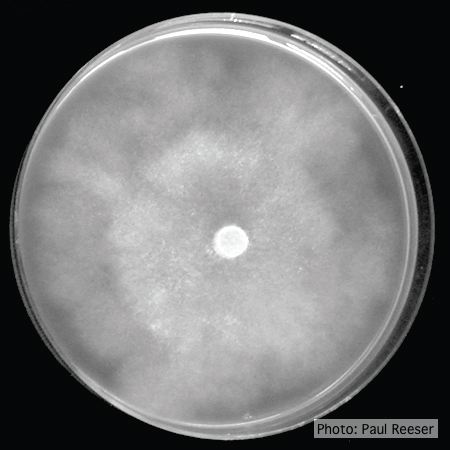 P. pseudotsugae colony growth on V8 agar |
|
P. kernoviae canker  Bole lesion on Fagus sylvatica |
P. pinifolia hyphal growth 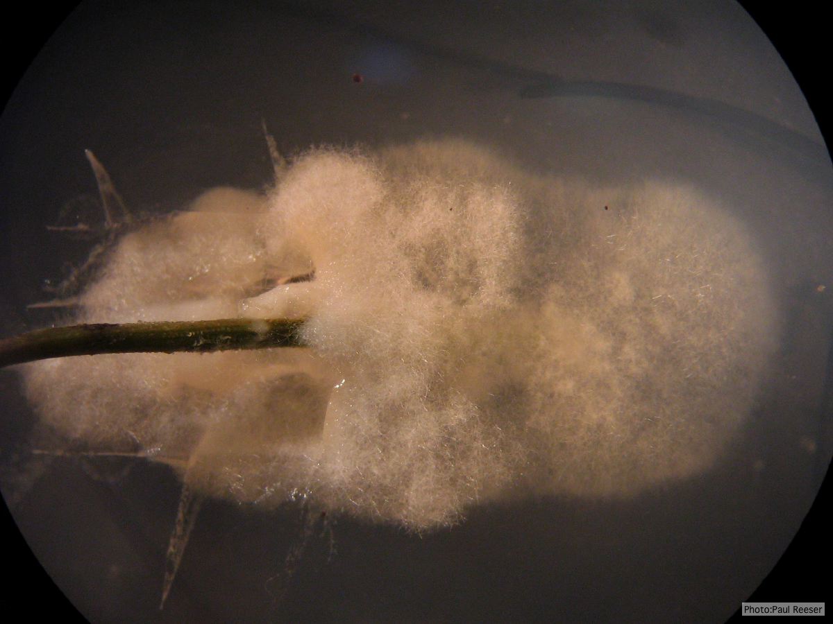 P. pinifolia pathogen growing from infected needle on selective agar |
Growth morphology on V8 of P. lateralis 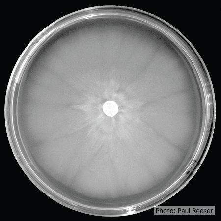 Colony morphology on V8 at 14 days |
|
P. pluvialis symptoms 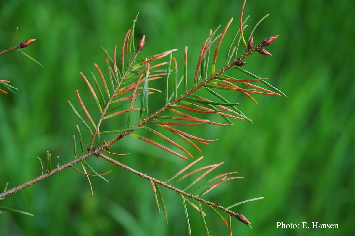 Symptoms of red needle cast on Douglas-fir needles |
P. boehmeriae oogonium 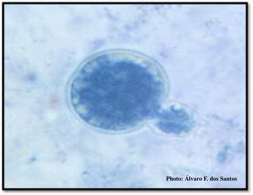 Oogonia and oospores with amphigynous antheridia |
P. cambivora sporangia 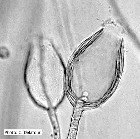 Empty sporangia of P. cambivora showing nested internal proliferation |
|
P. chlamydospora chlamydospore 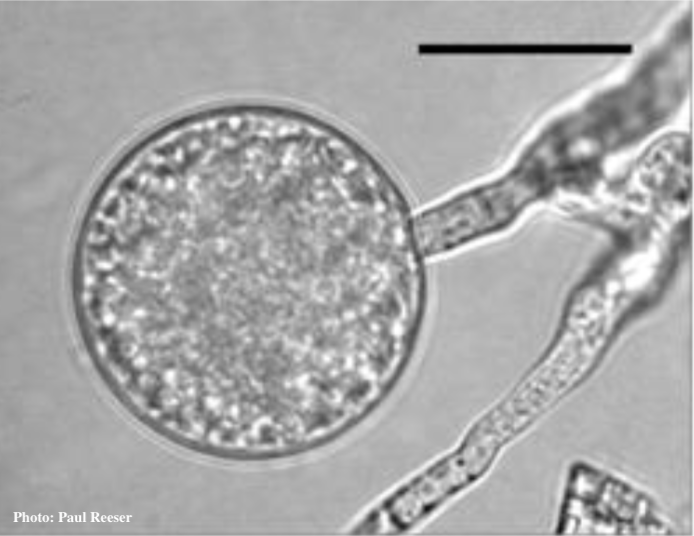 Phytophthora chlamydospora chlamydospore in agar. Bar is 20 µm. |
P. cactorum bleeding canker 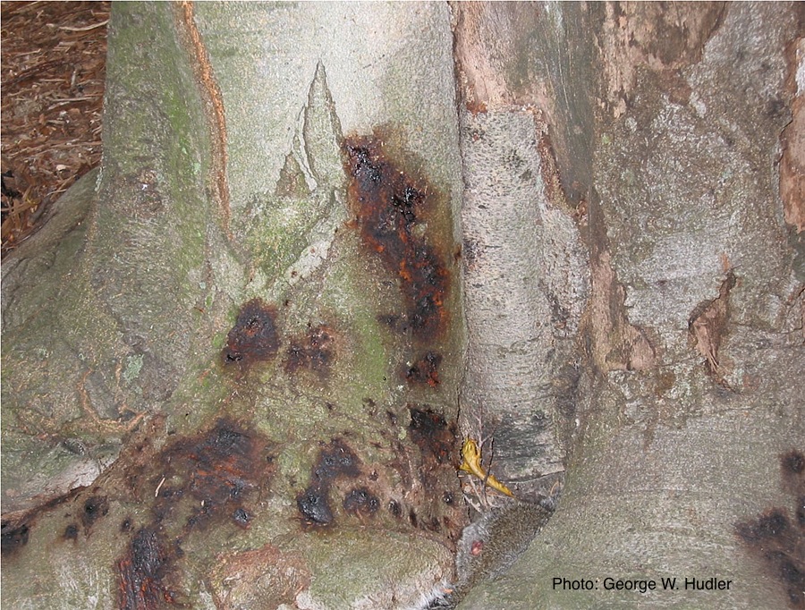 Bleeding canker on European beech (Fagus sylvatica) |
P. kernoviae disease on beech 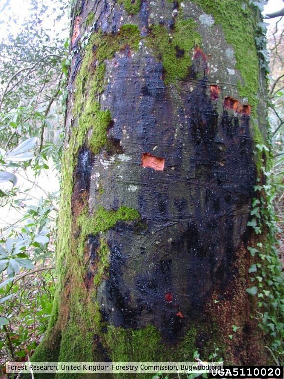 External lesion; 14 November 2003 |