

Symptoms of gummosis on black wattle (Fitopatol. bras. 2005)
Photo Gallery
|
P. nicotianae symptoms 2  |
P. kernoviae colony morphology on V8 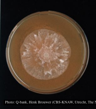 Colony morphology at 7 days at 18°C on V8, photo from Q-bank, used with permission. |
P. megasperma sporangia 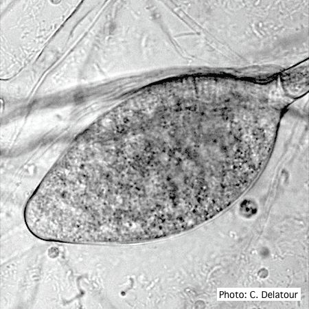 Ovoid, non-papillate sporangia showing internal proliferation of sporangiophore |
|
P. ramorum sporangium 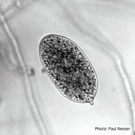 P. ramorum sporangium |
P. pinifolia on Pinus radiata 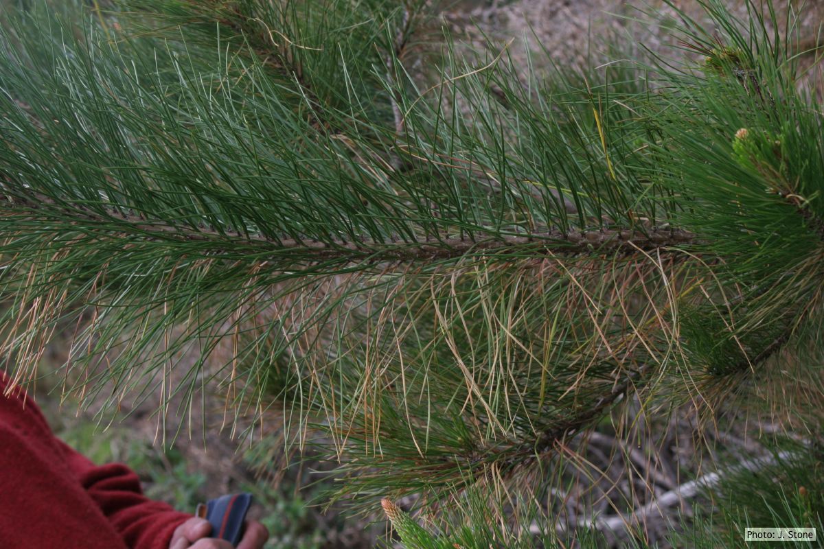 Dead needles on lower side of P. radiata branch. |
P. pluvialis on Pinus radiata in New Zealand  Pinus radiata needles showing colour changes following infection with red needle cast disease. The tissues around the initial infection at the base or along the needle senesce, and change yellow and then brown as indicated by the arrows before the needles cast. |
|
P. kernoviae oogonium 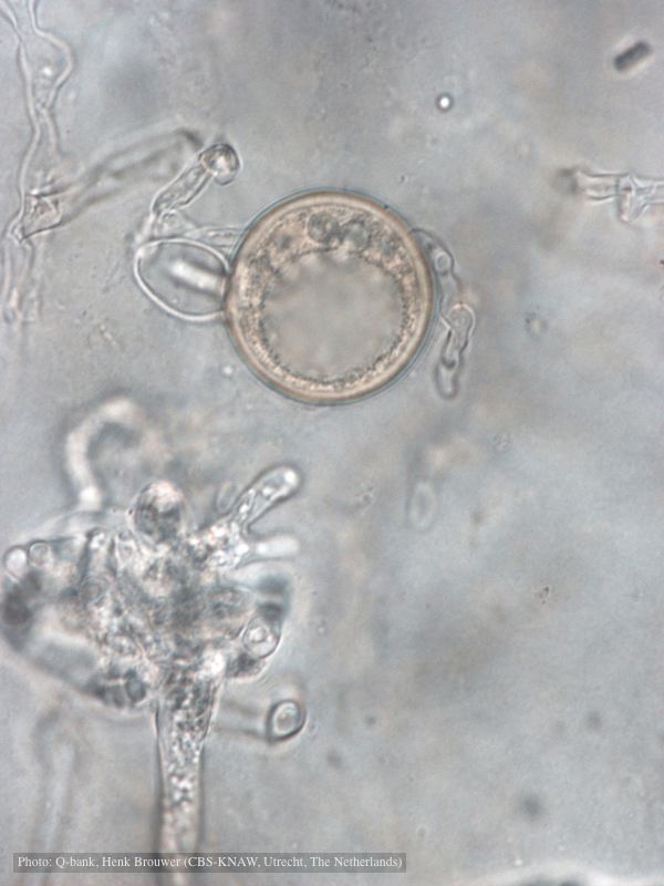 Oogonium with amphigynous antheridia, photo from Q-bank, used with permission |
P. pinifolia coenocytic hyphae 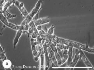 Coenocytic hyphae (from Duran et al. 2008). Scale bar = 20 μm. |
P. cinnamomi colony morphology on V8 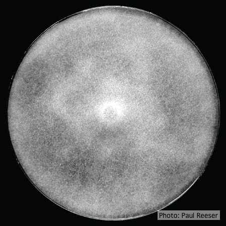 P. cinnamomi colony growth on V8 at 14 days |
|
P. cambivora disease symptoms 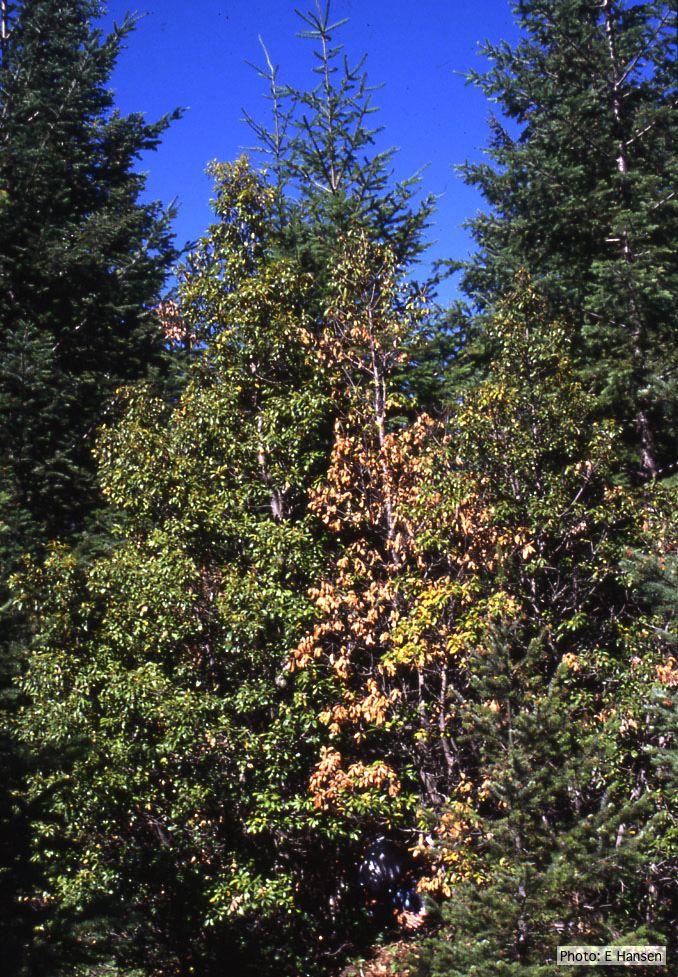 Dead and dying chinquapin infected with P. cambivora |
P. agathidicida growth on CMA 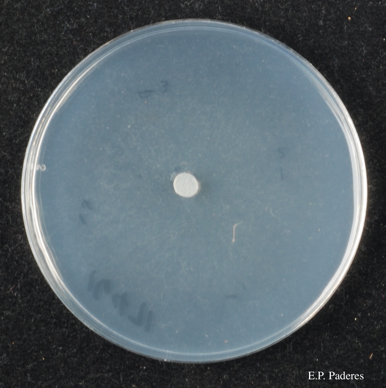 Diffuse, non-patterned, colony morphology of ICMP 16471 (the original “Gadgil isolate”) after 10-days incubation at 20°C in the dark |
P. cactorum bleeding canker  Bleeding canker on European beech (Fagus sylvatica) |