

Dead and dying chinquapin infected with P. cambivora
Photo Gallery
|
P. cambivora on dead and dying chinquapin  |
P. alni lesion in alder, Illwald, France 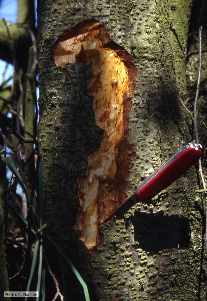 P. alni lesion in alder, Illwald, France |
P. pluvialis colony morphology on carrot agar  Colony morphology on carrot agar at 20 days |
|
P. katsurae sporangia 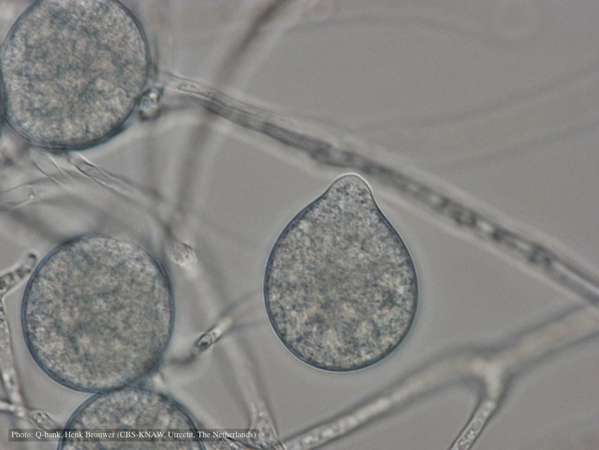 Papillate, non-caducous sporangia; photo used with permission from Q-bank |
P. siskiyouensis bleeding canker 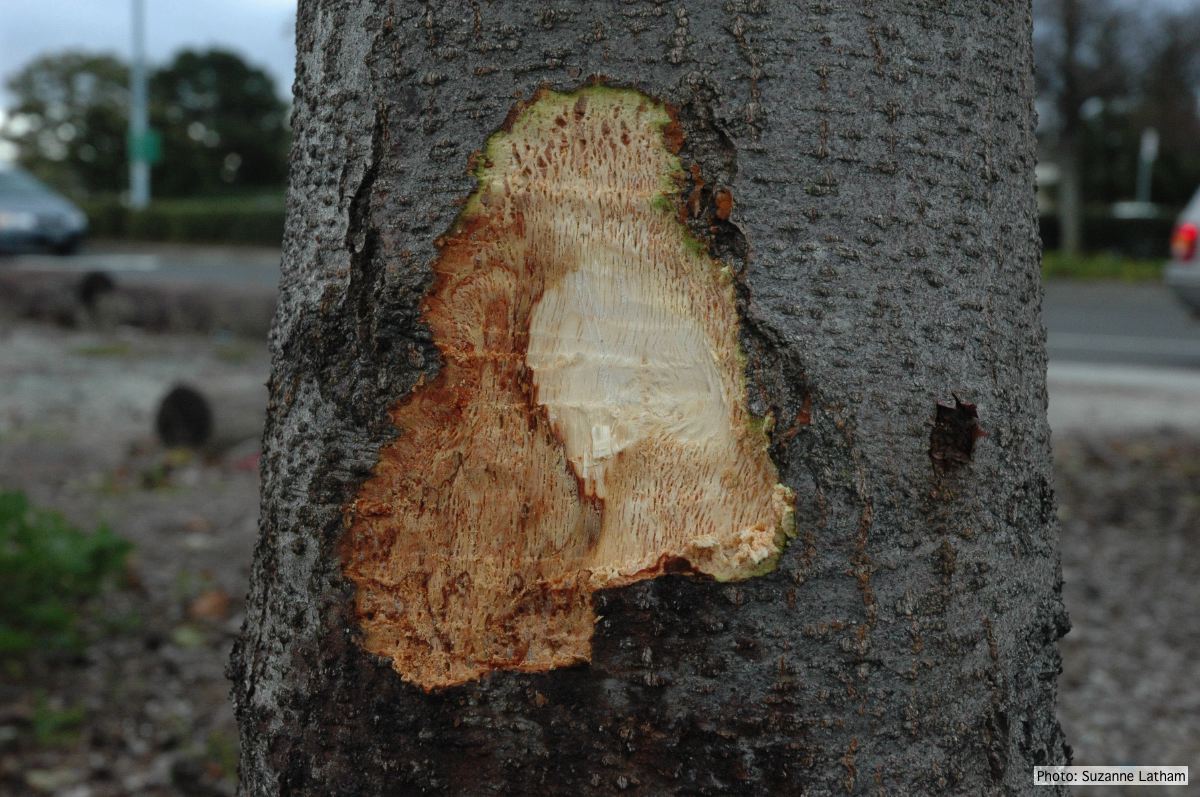 Close-up of margin area of bole lesions under the bark of a bleeding canker |
Comparative gametangial morphology of Phytophthora Clade 5 species  Comparative gametangial morphology of Phytophthora Clade 5 species, with SEM (top) and light microscopy (bottom). P. heveae has smooth walled oogonia with funnel-shaped, amphigynous antheridia. P. agathidicida has mildly stipulate oogonia with globose amphigynous antheridia. P.cocois has mildly bullate oogonia with reflexed amphigynous antheridia. P. castaneae has coarsely bullate oogonium with rugose protuberances and narrow amphigynous antheridia (Weir et al. 2015). |
|
P. palmivora oogonium 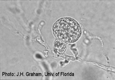 P. palmivora oogonium |
P. pseudosyringae hyphal swellings  Sub-globose hyphal swellings in water |
P. austrocedrae - Mal del ciprés, stages of decline 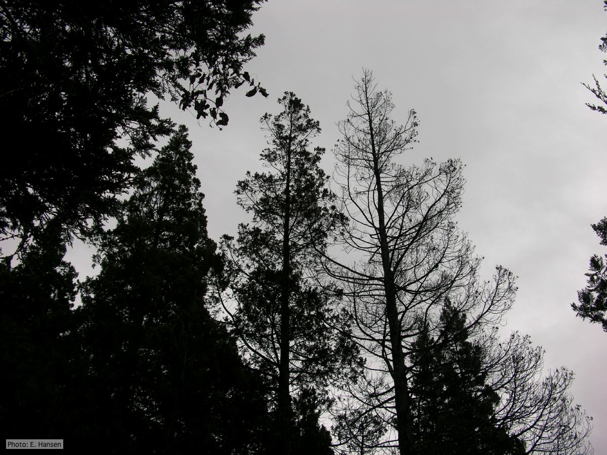 Mal del ciprés, stages of decline |
|
Vehicle washing  Truck washing to avoid spread of P. lateralis |
Growth of P. palmivora on CMA 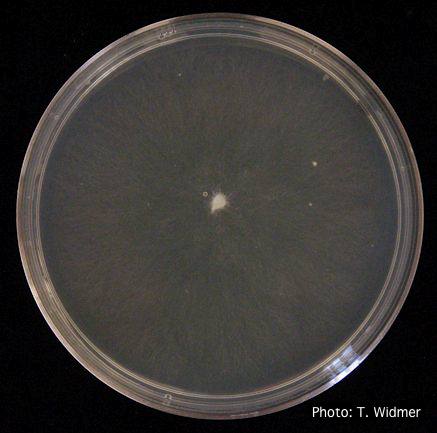 Growth of P. palmivora on corn meal agar |
P. agathadicida disease symptom 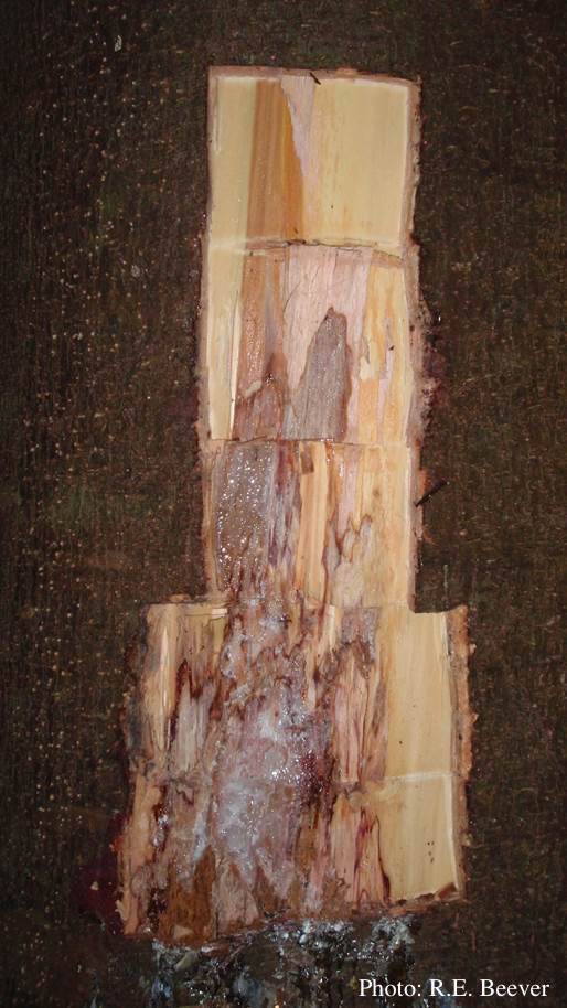 Excavated lesion, with outer bark removed showing extent of disease-front |