

P. chlamydospora colony morphology on V8 agar
Photo Gallery
|
P. chlamydospora colony morphology on V8 agar 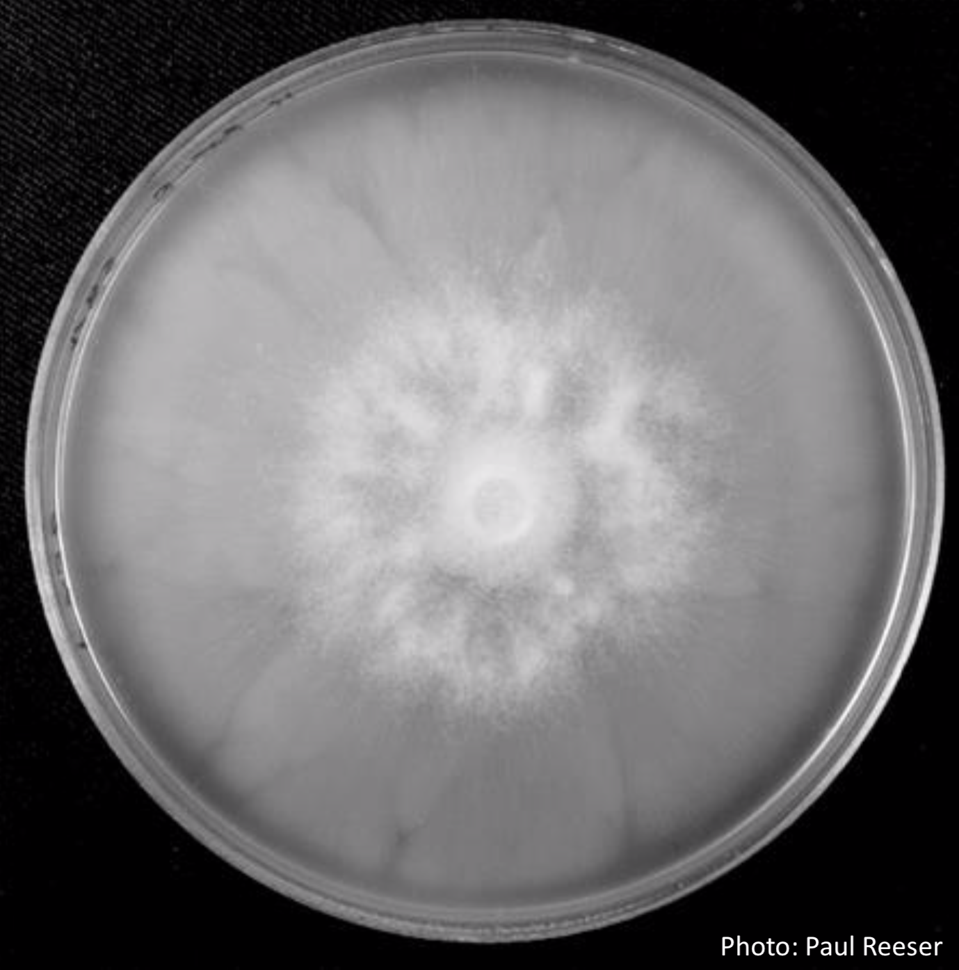 |
P. kernoviae sporangium 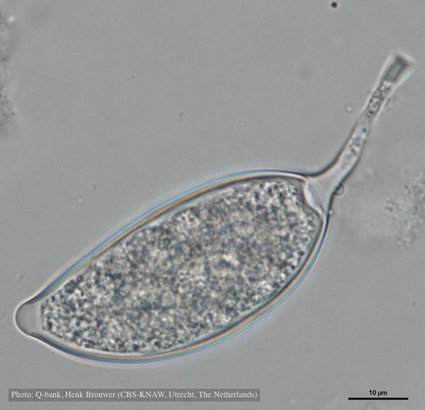 Asymmetrical sporangium, photo from Q-bank, used with permission |
P. cactorum bleeding canker 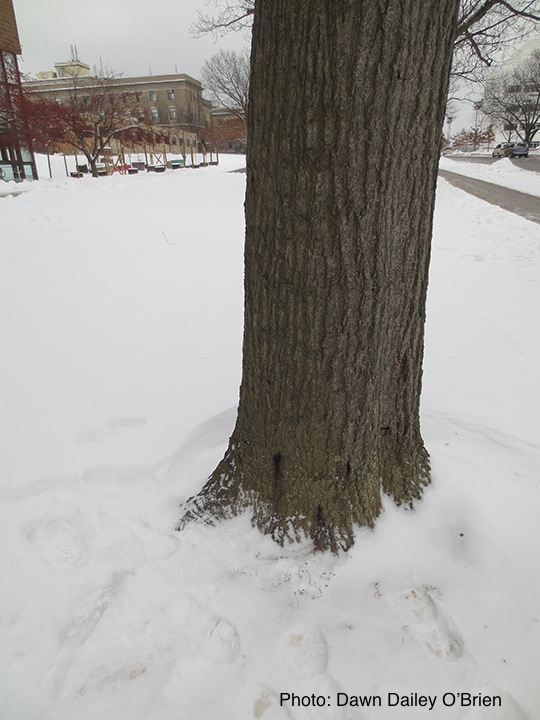 Bleeding canker on red oak (Quercus rubra) |
|
P. cambivora sporangium 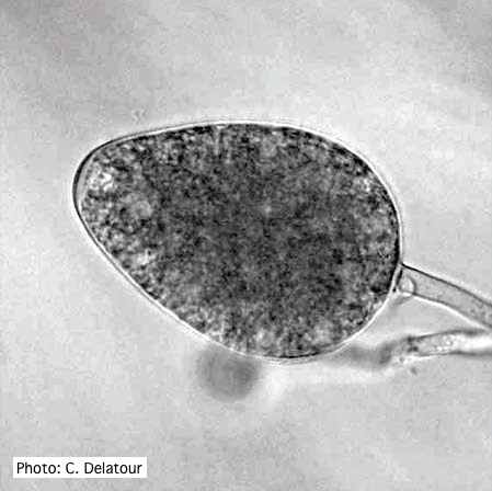 Ovoid non- papillate sporangia |
P. katsurae disease symptoms  Infected chestnut tree with girdling canker on stem |
P. nemorosa sporangium  Ovoid, semi-papillate sporangium showing medium length pedicel. |
|
Dead and healthy Port-Orford cedar seedlings  Port-Orford-cedar seedlings planted to test for Phytophthora lateralis resistance at the Dorena Genetic Resource Center |
P. alni oogonia subspecies and variants 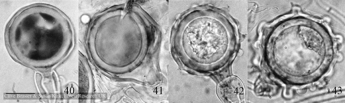 Fig. 40. P. alni subsp. uniformis. Fig. 41. P. alni subsp. multiformis German variant. Fig. 42. P. alni subsp. alni. Fig. 43. |
P. ramorum colony morphology on PDA 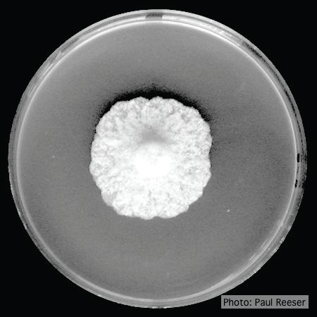 P. ramorum colony morphology on PDA |
|
P. arenaria sporangia  Globose papillate sporangia of Phytophthora arenaria on V8 agar flooded with soil extract. (Scale bar = 20 μm) |
P. lateralis on Port Orford cedar  Root lesions on Chaemacyparis lawsoniana |
Sporangia showing ovoid and ovoid to spherical shape and papillate condition sporangia  Sporangia showing ovoid and ovoid to spherical shape and papillate condition |